. 2019 Mar 20;9(1):4907.
doi: 10.1038/s41598-019-41373-0.

Noninvasive Contrast-Free 3D Evaluation of Tumor Angiogenesis with Ultrasensitive Ultrasound Microvessel Imaging

Affiliations

Noninvasive Contrast-Free 3D Evaluation of Tumor Angiogenesis with Ultrasensitive Ultrasound Microvessel Imaging

Chengwu Huang et al. Sci Rep. .

Abstract

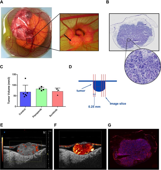
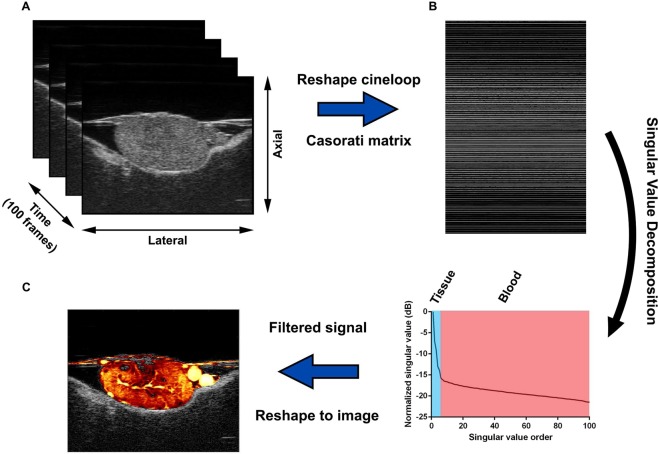
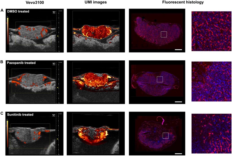
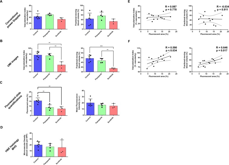
Ultrasound microvessel imaging (UMI), when applied with ultrafast planewave acquisitions, has demonstrated superior blood signal sensitivity in comparison to conventional Doppler imaging. Here we propose a high spatial resolution and ultra-sensitive UMI that is based on conventional line-by-line high-frequency ultrasound imagers and singular value decomposition (SVD) clutter filtering for the visualization and quantification of tumor microvasculature and perfusion. The technique was applied to a chicken embryo tumor model of renal cell carcinoma that was treated with two FDA-approved anti-angiogenic agents at clinically relevant dosages. We demonstrate the feasibility of 3D evaluation with UMI to achieve highly sensitive detection of microvasculature using conventional line-by-line ultrasound imaging on a preclinical and commercially available high-frequency ultrasound device without software or hardware modifications. Quantitative parameters (vascularization index and fractional moving blood volume) derived from UMI images provide significantly improved evaluation of anti-angiogenic therapy response as compared with conventional power Doppler imaging, using histological analysis and immunohistochemistry as the reference standard. This proof-of-concept study demonstrates that high-frequency UMI is a low-cost, contrast-agent-free, easily applicable, accessible, and quantitative imaging tool for tumor characterization, which may be very useful for preclinical evaluation and longitudinal monitoring of anti-cancer treatment.


Conflict of interest statement

The authors declare no competing interests.

Figures

Figure 1
Figure 1
Functional imaging of CAM engrafted cell-line tumors. (A) Overhead view of an ex ovo chicken embryo model. Cancer cell lines engrafted into the CAM resulted in highly vascularized, solid, and spheroidal tumor masses. (B) H&E section taken from representative control tumor (DMSO treated). High magnification section reveals viable tumor nuclei with a high degree of adjacent vasculature. (C) Endpoint tumor volumes for each treatment group. Differences in volume were not found to be significant. Data are mean ± s.d. (D) Schematic representation of tumor imaging protocol. Tumors were imaged throughout their volume with a step size of 0.25 mm. (E) Representative image of control (DMSO) treated Renca tumor obtained with conventional power Doppler of VisualSonics Vevo® 3100. (F) SVD-based clutter filtering of ultrasound echoes from the tumor in (E) resulted in improved vascular imaging (Supplementary Video S1). (G) Fluorescent histology section of the example tumor, which confirms high intratumoral vascularization (red signal).
Figure 2
Figure 2
SVD-based clutter filtering of Vevo® 3100 cineloops demonstrated superior microvascular detection and reduced tissue background. (A) B-mode IQ cineloops (100 frames) were considered as three-dimensional imaging volumes with axial, lateral, and slow-time dimensions. (B) These spatiotemporal data sets were reshaped into 2D Casorati matrices in a column-wise manner. Singular value decomposition of Casorati matrices decomposed the data into singular vectors and singular values, allowing for the extraction of blood signals. (C) An inverse SVD was performed to recover blood flow signals back to the spatiotemporal domain, resulting in a superior microvascular image in comparison to conventional power Doppler mode.
Figure 3
Figure 3
UMI improved detection and quantification of anti-angiogenic treatment effect. (A) Control tumor exhibited a high degree of intratumoral vascularization in conventional power Doppler imaging and UMI (Supplementary Video S2). H&E histology confirmed viable tumor cells and the presence of dense capillary networks. Fluorescent histology permitted the quantification of MFI and fluorescent vessel area as surrogate measures of microvascular density. (B) Conventional power Doppler imaging of pazopanib treated tumor did not reveal a significant change in vascularization. UMI image revealed localized avascular regions. Histology revealed some regional necrosis and a slightly reduced microvascular density (Supplementary Video S3). (C) Sunitinib treated tumor exhibited a modest decrease in tumor vascularization on Vevo®’s conventional Doppler image. This anti-angiogenic effect was more pronounced in UMI image (Supplementary Video S4). Histology confirmed intratumoral necrosis and a reduced microvascular density. Scale bar is 1 mm. Refer to Supplementary Fig. S1 and supplemental videos for 3D images. Rotation animations of the volumetric renderings are displayed in Supplementary Video S5.
Figure 4
Figure 4
Summary statistics confirmed anti-angiogenic treatment effect. (A) Conventional power Doppler imaging using the Vevo® 3100 system did not detect a significant decrease in intratumoral vasculature due to anti-angiogenic therapy. (B) The proposed UMI method based on the B-mode IQ data detected a significant decrease in vascularization index (VI) for sunitinib (multiplicity adjusted p = 0.0017) treated tumors, but not for pazopanib (p = 0.7251), in comparison to control. The confidence of this result improved when using fractional moving blood volume (FMBV) images for analysis (p = 0.0005). In both cases, pazopanib treated tumors were also found to be significantly different from the sunitinib treated cohort (p = 0.0048 for VI, and p = 0.0054 for FMBV). (C) Quantifications of fluorescent histology confirmed an anti-angiogenic treatment effect for both therapies (p = 0.0065 for pazopanib, and p = 0.0034 for sunitinib). (D) Microvascular density (MVD) evaluated from H&E slides did not demonstrate a significant therapy effect (p = 0.7364 and p = 0.6382 for pazopanib and sunitinib, respectively). (E) Conventional power Doppler imaging did not show a significant correlation for VI (p = 0.778) or FMBV (p = 0.911). (F) For UMI images, both VI and FMBV were moderately correlated with fluorescent area percentage (R = 0.590, p = 0.034 and R = 0.646, p = 0.017, respectively). All data are mean ± s.d. P-values are from a one-way ANOVA using the Holm-Sidak test to correct for multiple comparisons, and are reported as multiplicity adjusted values.

Similar articles

Cited by

References

    1. Michiels C. Physiological and Pathological Responses to Hypoxia. Am. J. Pathol. 2004;164:1875–1882. doi: 10.1016/S0002-9440(10)63747-9. - DOI - PMC - PubMed
    1. Carmeliet P, Jain RK. Angiogenesis in cancer and other diseases. Nature. 2000;407:249–257. doi: 10.1038/35025220. - DOI - PubMed
    1. Siemann DW. The Unique Characteristics of Tumor Vasculature and Preclinical Evidence for its Selective Disruption by Tumor-Vascular Disrupting Agents. Cancer Treat. Rev. 2011;37:63–74. doi: 10.1016/j.ctrv.2010.05.001. - DOI - PMC - PubMed
    1. Carmeliet P. Angiogenesis in life, disease and medicine. Nature. 2005;438:932–936. doi: 10.1038/nature04478. - DOI - PubMed
    1. Goel S, et al. Normalization of the vasculature for treatment of cancer and other diseases. Physiol. Rev. 2011;91:1071–1121. doi: 10.1152/physrev.00038.2010. - DOI - PMC - PubMed

Publication types

MeSH terms