. 2019 Mar 20;9(1):4916.
doi: 10.1038/s41598-019-41471-z.

Nerve electrical stimulation enhances osseointegration of implants in the beagle

Affiliations

Nerve electrical stimulation enhances osseointegration of implants in the beagle

Ping Zhou et al. Sci Rep. .

Abstract

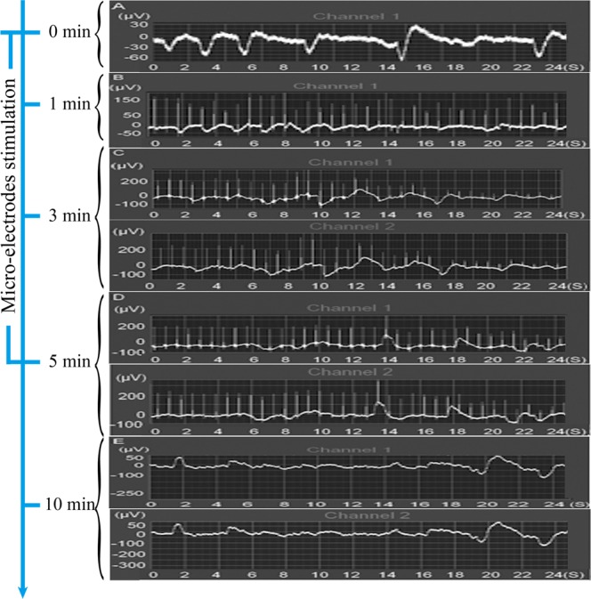
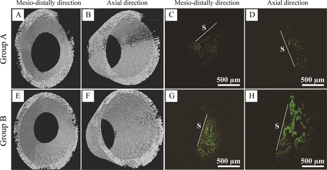
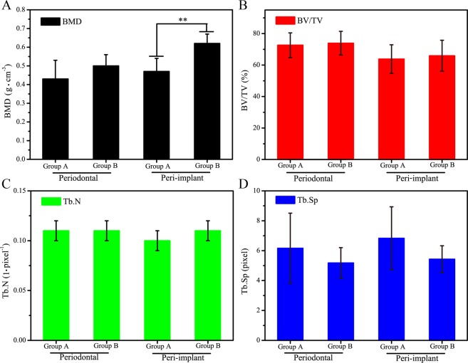
Dental implantation has been the primary method for the treatment of tooth loss, but longer than 3 months healing times are generally required. Because immediate load implants are suitable only for certain categories of implant patients, it has value to develop a novel method to facilitate the implant-bone osseointegration process. Cylindrical titanium implants were implanted in the tooth sockets of beagles, and microelectrode stimulation of the sympathetic nerves in the infraorbital nerve was performed after implantation for 1 week. The authors found that one-sided nerve stimulation was shown to evoke consistent electric potential changes in both sides of the infraorbital nerves. Moreover, after 4 weeks of implantation, more new bone was clearly observed around the implants in the beagles that received electrical stimulation treatment than was observed in the control animals. Furthermore, a higher mineralization density was measured in the new peri-implant bone tissues of the stimulated beagles when compared to controls. These results demonstrate that the simple and safe physical method of microelectrode stimulation to sympathetic nerves can promote the formation of new bone and the osseointegration of implants. This technique is worth promoting and has the potential to reduce the healing time of dental implantation in future clinical cases.


Conflict of interest statement

The authors declare no competing interests.

Figures

Figure 1
Figure 1
The electric potential of sympathetic nerve fibers in the infraorbital nerve of beagles before (A) and after microelectrode stimulation for various times (1 min, 3 min and 5 min) (B–D) was measured in real time by a multiple physiologic recorder. The stimulation was stopped for 5 min, and the electric potential was detected again. (E) In this figure, channel 1 and channel 2 were applied to detect the electric potential of the left and right sympathetic nerve fibers, respectively.
Figure 2
Figure 2
Microelectrode stimulation promotes the osseointegration of implants in beagles. Representative micro-CT images for implants of group A (A) and group B (B) in the horizontal direction. Group B animals were stimulated by microelectrodes daily for 3 weeks, while group A animals received no treatments and served as controls. The white arrows indicate the gaps between the implants and the surrounding bone tissue.
Figure 3
Figure 3
Microelectrodes stimulation facilitated new bone formation. Representative radiographs of micro-CT 3D reconstruction models for surrounding bone of group A (A,B) and group B (E,F) implants after implantation for 4 weeks. The new bone formation around implants of group A (C,D) and group B (G,H) were examined using bone labeling. Calcein (green) and tetracycline hydrochloride (yellow) were injected on weeks 2 and 4, respectively. White lines indicate the interface between the samples (S) and the new bone. Scale bars, 500 µm.
Figure 4
Figure 4
Comparisons of the indicated parameters of periodontal and peri-implant bone between group (A) and group (B) animals. All measurements were obtained from a micro-CT after implantation for 4 weeks. BMD represents bone mineral density (A); BV/TV represents percent bone volume (B); Tb.N represents trabecular number (C); Tb.Sp represents trabecular spacing (D). n = 7, **represents p < 0.01.

Similar articles

Cited by

References

    1. Binon PP. Implants and components: entering the new millennium. Int. J. Oral Maxillofac. Implants. 2000;15:76–94. - PubMed
    1. Puleo DA, Nanci A. Understanding and controlling the bone–implant interface. Biomaterials. 1999;20:2311–2321. doi: 10.1016/S0142-9612(99)00160-X. - DOI - PubMed
    1. De GK, Wolke JG, Jansen JA. Calcium phosphate coatings for medical implants. Proceedings of the Institution of Mechanical Engineers Part H Journal of Engineering in Medicine. 1998;212:137–147. doi: 10.1243/0954411981533917. - DOI - PubMed
    1. Bernhardt R, et al. Osteoconductive modifications of Ti-implants in a goat defect model: characterization of bone growth with SR muCT and histology. Biomaterials. 2005;26:3009–3019. doi: 10.1016/j.biomaterials.2004.08.030. - DOI - PubMed
    1. Le GL, Soueidan A, Layrolle P, Amouriq Y. Surface treatments of titanium dental implants for rapid osseointegration. Dent. Mater. 2007;23:844–854. doi: 10.1016/j.dental.2006.06.025. - DOI - PubMed

Publication types

MeSH terms