. 2019 Apr;568(7752):420-423.
doi: 10.1038/s41586-019-1026-5. Epub 2019 Mar 20.

Novel tau filament fold in chronic traumatic encephalopathy encloses hydrophobic molecules

Affiliations

Novel tau filament fold in chronic traumatic encephalopathy encloses hydrophobic molecules

Benjamin Falcon et al. Nature. 2019 Apr.

Abstract

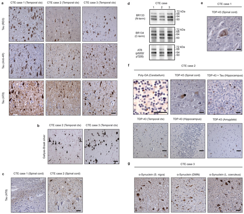
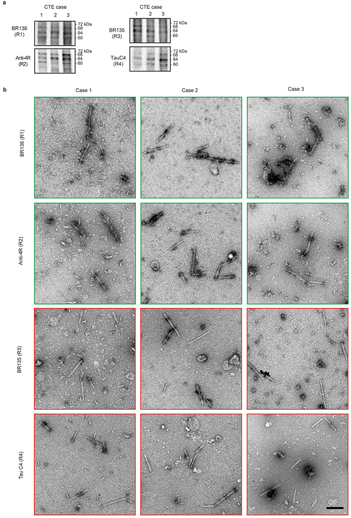
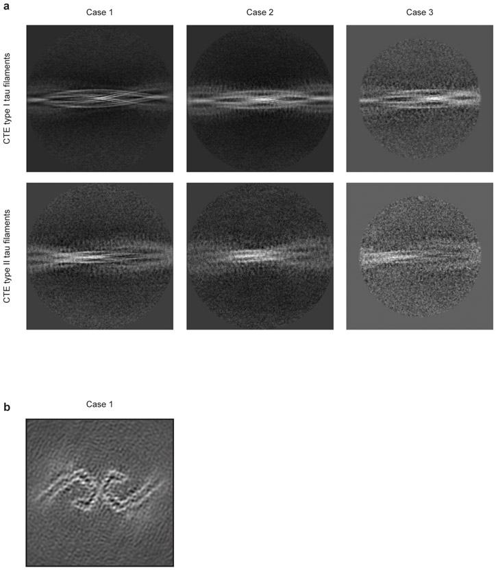
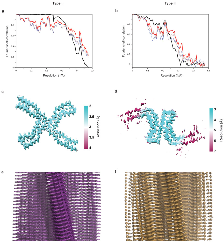
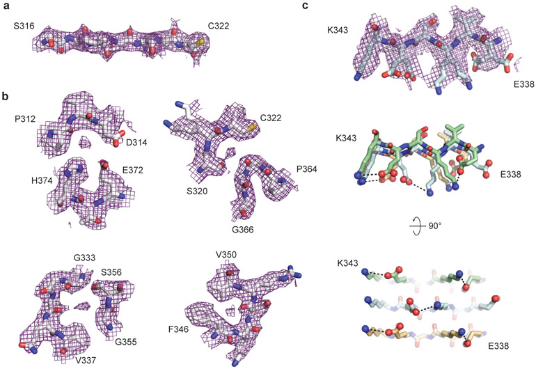
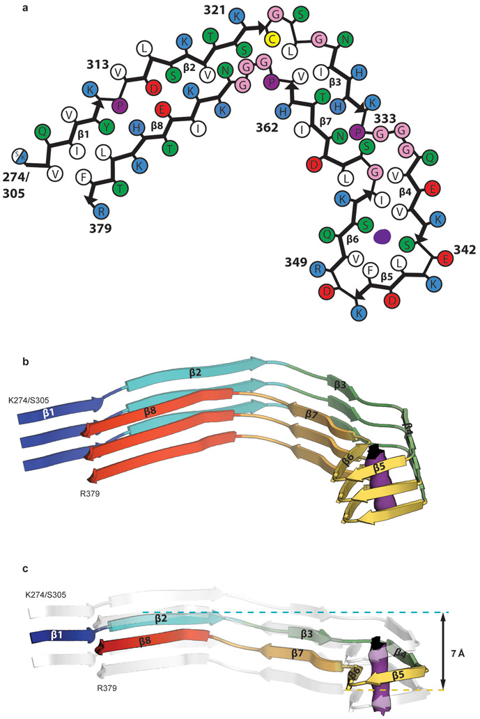
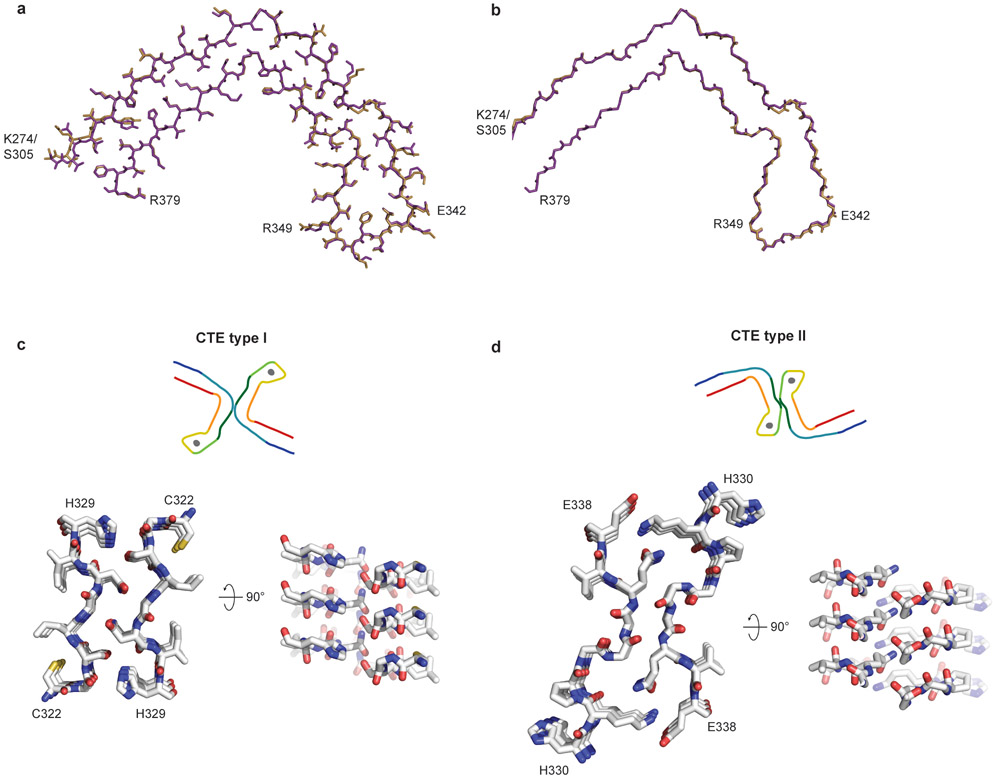
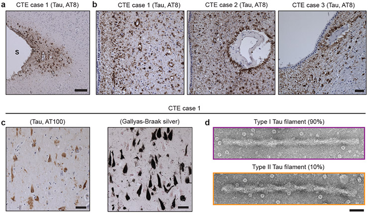
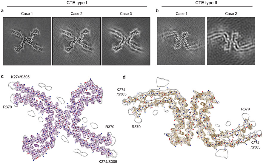
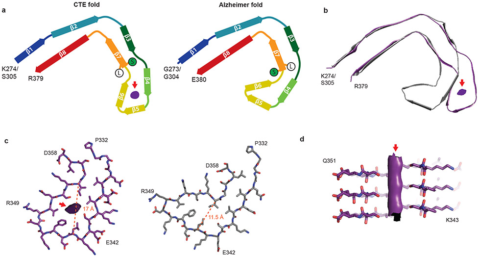
Chronic traumatic encephalopathy (CTE) is a neurodegenerative tauopathy that is associated with repetitive head impacts or exposure to blast waves. First described as punch-drunk syndrome and dementia pugilistica in retired boxers1-3, CTE has since been identified in former participants of other contact sports, ex-military personnel and after physical abuse4-7. No disease-modifying therapies currently exist, and diagnosis requires an autopsy. CTE is defined by an abundance of hyperphosphorylated tau protein in neurons, astrocytes and cell processes around blood vessels8,9. This, together with the accumulation of tau inclusions in cortical layers II and III, distinguishes CTE from Alzheimer's disease and other tauopathies10,11. However, the morphologies of tau filaments in CTE and the mechanisms by which brain trauma can lead to their formation are unknown. Here we determine the structures of tau filaments from the brains of three individuals with CTE at resolutions down to 2.3 Å, using cryo-electron microscopy. We show that filament structures are identical in the three cases but are distinct from those of Alzheimer's and Pick's diseases, and from those formed in vitro12-15. Similar to Alzheimer's disease12,14,16-18, all six brain tau isoforms assemble into filaments in CTE, and residues K274-R379 of three-repeat tau and S305-R379 of four-repeat tau form the ordered core of two identical C-shaped protofilaments. However, a different conformation of the β-helix region creates a hydrophobic cavity that is absent in tau filaments from the brains of patients with Alzheimer's disease. This cavity encloses an additional density that is not connected to tau, which suggests that the incorporation of cofactors may have a role in tau aggregation in CTE. Moreover, filaments in CTE have distinct protofilament interfaces to those of Alzheimer's disease. Our structures provide a unifying neuropathological criterion for CTE, and support the hypothesis that the formation and propagation of distinct conformers of assembled tau underlie different neurodegenerative diseases.


Figures

Extended Data Figure 1:
Extended Data Figure 1:. Further characterization of tau pathologies in CTE cases 1-3. Coexisting pathologies.
a, Staining of tau inclusions in the temporal cortex of CTE cases 1, 2 and 3 using antibodies RD3 (3R tau), Anti-4R (4R tau) and AT8 (pS202/pT205 tau). Scale bar, 50 μm. b, Gallyas-Braak silver staining of tau inclusions in the temporal cortex of CTE cases 2 and 3. Scale bar, 50 μm. c, Staining of tau inclusions in the spinal cord of CTE cases 1 and 2 using antibody AT8. Scale bar, 50 μm. d, Immunoblots of sarkosyl-insoluble tau from the temporal cortices of CTE cases 1, 2 and 3 using antibodies BR133, BR134 and AT8. e, Staining of a TDP-43 inclusion in the spinal cord of CTE case 1. Scale bar, 25 μm. f, Staining of CTE case 2 for poly-GA inclusions in the cerebellum and TDP-43 inclusions in the spinal cord, temporal cortex, hippocampus and amygdala. In the top right-hand panel, double-labelling of tau inclusions (AT8; brown) and TDP-43 inclusions (red) is shown. Scale bars, 25 μm (upper panels) and 50 μm (lower panels). g, Staining of CTE case 3 for α-synuclein inclusions in the substantia nigra, dorsal motor nucleus (DMN) of the vagus nerve and locus coeruleus. Scale bar, 50 μm. Nuclei were counterstained blue in all images.
Extended Data Figure 2:
Extended Data Figure 2:. Immunolabelling of tau filaments extracted from the brains of CTE cases 1-3.
a,b, Immunoblots and immunolabelling of tau filaments extracted from the temporal cortices of CTE cases 1, 2 and 3. a, Immunoblots of sarkosyl-insoluble tau using antibodies BR136, Anti-4R, BR135 and TauC4. b, Representative immuno-EM images of tau filaments labelled with antibodies BR136 and Anti-4R. Unlike BR136 and Anti-4R, antibodies BR135 and TauC4 did not label the filaments, indicating that their epitopes lie within the ordered filament cores. Scale bar, 200 nm.
Extended Data Figure 3:
Extended Data Figure 3:. Cryo-EM two-dimensional and three-dimensional classification.
a, 2D-class averages spanning an entire helical crossover of type I and type II tau filaments from CTE cases 1-3. b, Cryo-EM structure of PHFs from the temporal cortex of CTE case 1.
Extended Data Figure 4:
Extended Data Figure 4:. Cryo-EM map and model comparisons.
a,b, Fourier shell correlation curves between two independently refined half-maps (bold black line); between the final cryo-EM reconstruction and refined atomic model (bold red line); between the first half-map and the atomic model refined against the first half map (brown line); and between the second half map and the atomic model refined against the first half map (blue dashed line) for CTE type I (a) and type II (b) filaments. c,d, Local resolution estimates for the CTE type I (c) and type II (d) filament reconstructions. e,f, Views normal to the helical axis of the CTE type I (e) and type II (f) filament reconstructions.
Extended Data Figure 5:
Extended Data Figure 5:. Cryo-EM map of type I tau filaments from CTE case 1.
a-c, Close-up view of the cryo-EM map with the atomic model overlaid showing densities for oxygens atoms of the peptide groups (a) and ordered solvent molecules (b). c, Side-chain conformations for the alternating positively and negatively charged solvent-exposed side-chains of residues 336-343 in successive rungs.
Extended Data Figure 6:
Extended Data Figure 6:. CTE tau filament fold.
a, Schematic view of the CTE tau filament fold. b, Rendered view of the secondary structure elements in the CTE fold, depicted as three successive rungs. c, As in b, but in a view perpendicular to the helical axis, revealing the changes in height within a single molecule.
Extended Data Figure 7:
Extended Data Figure 7:. Comparison of CTE type I and type II tau filament folds and protofilament interfaces.
a, The type I protofilament structure is shown in purple and the type II protofilament structure in gold. b, As in a, but showing backbone atoms only. c,d, Packing between residues 322CGSLGNIH329 of the two protofilaments in type I filaments (c) and between residues 331KPGGGQVE338 of the two protofilaments in type II filaments (d).
Extended Data Figure 8:
Extended Data Figure 8:. Measurement and modelling of optical aberrations.
Three-fold astigmatism (antisymmetrical; upper panels) and 4-fold astigmatism (symmetrical; lower panels) measured in per-Fourier-pixel average phase-error plots (left images) and represented by our parametric model (right images) for CTE type I tau filaments from case 1. The plot shows image frequencies up to 2.9 Å. The outlier ring at 4.7 Å corresponds to the dominant frequency given by a double-repetition of the helix. This ring has been excluded from the parametric fit.
Figure 1:
Figure 1:. Filamentous tau pathology of CTE.
a, Perivascular staining of tau inclusions in the depth of a sulcus (S) in the frontal cortex of CTE case 1 using antibody AT8 (pS202/pT205 tau; brown). Nuclei were counterstained blue. Scale bar, 100 μm. b, Perivascular staining of tau inclusions in the subependymal regions of CTE cases 1, 2 and 3 using antibody AT8 (brown). Nuclei were counterstained blue. Scale bar, 100 μm. c, Staining of tau inclusions using antibody AT100 (pT212/pS214/pT217 tau; brown) and Gallyas-Braak silver staining (black) of inclusions in the temporal cortex of CTE case 1. Nuclei were counterstained blue. Scale bar, 50 μm. d, Negative-stain electron micrographs of a type I and a type II tau filament extracted from temporal cortex of CTE case 1. Scale bar, 50 nm.
Figure 2:
Figure 2:. Cryo-EM structures of CTE type I and type II tau filaments.
a,b, Cryo-EM structures of CTE type I tau filaments from the temporal cortices of cases 1, 2 and 3 (a) and type II tau filaments from the temporal cortices of cases 1 and 2 (b). All structures show identical pairs of protofilaments and the same inter-protofilament packing between cases in CTE type I and type II filaments. c,d, Sharpened, high-resolution cryo-EM maps of CTE type I (c) and type II (d) tau filaments with their corresponding atomic models overlaid. Unsharpened, 4.5 Å low-pass filtered maps are shown as grey outlines, showing weaker densities extending from the N- and C-terminal regions, as well as bordering the solvent-exposed side-chains of K317 and K321, H362 and K369, and K369 and K375.
Figure 3:
Figure 3:. Comparison of the CTE and Alzheimer tau filament folds.
a, Schematic of the secondary structure elements in the CTE and Alzheimer folds, depicted as a single rung. The positions of S356 (green ‘S’) and L357 (white ‘L’) in the two folds are highlighted. The extra density is depicted in grey. b, Overlay of the CTE fold (purple) and the Alzheimer fold (grey), shown as a single rung. c, The β-helices of the Alzheimer fold (grey) and the CTE fold (purple), depicted as a single rung. The distances between the Cα atoms of L344 and I354 are shown as orange dashed lines. d, View normal to the helical axis of the CTE fold β-helix, depicted as three rungs and shown as a cross-section through S341 and S352.

Comment in

  • Distinct tau filaments in CTE.
    Kelsey R. Kelsey R. Nat Rev Neurol. 2019 Jun;15(6):308-309. doi: 10.1038/s41582-019-0185-1. Nat Rev Neurol. 2019. PMID: 30988502 No abstract available.

Similar articles

Cited by

References

    1. Martland HS Punch drunk. J. Am. Med. Assoc. 19, 1103–1107 (1928).
    1. Millspaugh JA Dementia pugilistica. US Nav Med. Bull. 35, 297–303 (1937).
    1. Corsellis JA, Bruton CJ & Freeman-Browne D The aftermath of boxing. Psychol. Med. 3, 270–303 (1973). - PubMed
    1. Omalu BI et al. Chronic traumatic encephalopathy in a National Football League player. Neurosurgery 57, 128–134 (2005). - PubMed
    1. McKee AC, Daneshvar DH, Alvarez VE & Stein TD The neuropathology of sport. Acta Neuropathol. 127, 29–51 (2014). - PMC - PubMed