. 2019 Mar 6:10:369.
doi: 10.3389/fimmu.2019.00369. eCollection 2019.

A Soluble Immune Effector Binds Both Fungi and Bacteria via Separate Functional Domains

Affiliations

A Soluble Immune Effector Binds Both Fungi and Bacteria via Separate Functional Domains

Assunta Liberti et al. Front Immunol. .

Abstract

The gut microbiome of animals consists of diverse microorganisms that include both prokaryotes and eukaryotes. Complex interactions occur among these inhabitants, as well as with the immune system of the host, and profoundly influence the overall health of both the host and its microbial symbionts. Despite the enormous importance for the host to regulate its gut microbiome, the extent to which animals generate immune-related molecules with the capacity to directly influence polymicrobial interactions remains unclear. The urochordate, Ciona robusta, is a model organism that has been adapted to experimental studies of host/microbiome interactions. Ciona variable-region containing chitin-binding proteins (VCBPs) are innate immune effectors, composed of immunoglobulin (Ig) variable regions and a chitin-binding domain (CBD) and are expressed in high abundance in the gut. It was previously shown that VCBP-C binds bacteria and influences both phagocytosis by granular amoebocytes and biofilm formation via its Ig domains. We show here that the CBD of VCBP-C independently recognizes chitin molecules present in the cell walls, sporangia (spore-forming bodies), and spores of a diverse set of filamentous fungi isolated from the gut of Ciona. To our knowledge, this is the first description of a secreted Ig-containing immune molecule with the capacity to directly promote transkingdom interactions through simultaneous binding by independent structural domains and could have broad implications in modulating the establishment, succession, and homeostasis of gut microbiomes.

Keywords: Ciona; VCBP-C; fungal-immune interaction; gut immunity; host-microorganism interactions; innate immunity; mycobiota; transkingdom interactions.


Figures
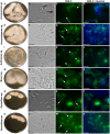
Figure 1
Figure 1
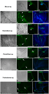
Immunofluorescence with the Fc-CBD-C probe on fungal sections. Several species of fungi, including: Acremonium sp., Acrostalagmus sp., Arthrinium sp., Mucor sp., and Penicillium spp., were isolated from Ciona gut and grown on YPD agar plates (A,E,I,M,Q,U). Immunofluorescence staining, using the Fc-CBD-C probe and detected with a fluorescent secondary antibody (Alexa Fluor 488), on OCT-embedded and cryo-sectioned fungal isolates, indicates that chitin molecules are distributed in the whole cell wall of most of the fungi analyzed (G,O,S,W, arrows). In two fungal species, the Fc-CBD-C probe detects chitin localized in specific regions of the fungal cell wall (C,K, arrowhead). (B,F,J,N,R,V) are brightfield images of fungal sections; (D,H,L,P,T,X) represents merged images of the Fc-CBD-C probe (green) and nuclear staining with Hoechst (blue). Scale bars: (B–D,J–L,R-T,V–X), 5 μm; (F–H, N–P) 10 μm.
Figure 2
Figure 2
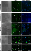
Immunofluorescence with the Fc-CBD-C probe on fungal sections. Fungal species, including Phaeosphaeria sp., Phoma sp., and Trichoderma spp., were isolated from the Ciona gut and grown on YPD agar plates (A,E,I,M). Immunofluorescent staining, using the Fc-CBD-C probe, on OCT-embedded and cryo-sectioned fungal isolates, indicates that chitin molecules are distributed in the whole cell walls of these fungi (C,G,K,O, arrows). (B,F,J,N) are brightfield images of fungal sections; (D,H,L,P) represent merged images of the Fc-CBD-C probe (green) and nuclear staining with Hoechst (blue). Scale bars: (B–D,J–L), 10 μm; (F–H,N–P), 5 μm.
Figure 3
Figure 3
Immunofluorescence with the Fc-CBD-C probe on whole fungi grown in liquid medium. Fungal species, Mucor sp. (A–F), Penicillium spp. (G–R), and Trichoderma sp. (S–X), were grown in liquid medium and characterized by immunofluorescent staining with the Fc-CBD-C probe. In Mucor sp., CBD-C binds fungal spores (B,E, arrowheads); in Penicillium spp., CBD-C recognizes chitin in specific regions of the hyphae (K,N,Q, arrows) and in some cases fungal spores (H, arrowhead); in Trichoderma sp., chitin fibers are apparent in specific regions of the hyphae (T,W, arrows). (A,D,G,J,M,P,S,V) represent brightfield images; (C,F,I,L,O,R,U,X) represents merged images of the Fc-CBD-C probe (green) and nuclear staining with Hoechst (blue). Scale bars: (A–C,D–F,J–L,P–R,V–X), 50 μm; (G–I,M–O,S–U), 25 μm.
Figure 4
Figure 4
Immunofluorescence with the Fc-CBD-C probe on fungal spores. In Acremonium sp., Acrostalagmus sp., and Mucor sp. the CBD-C probe detects chitin fibers in regionalized areas on the surface of spores isolated from liquid cultures (B,E,H, arrows). In Penicililum spp., CBD-C binds almost the entire spore wall (K,N, arrowheads) in most spores. In some spores, chitin is instead detected in localized spots (K,N, arrow). (A,D,G,J,M) represents brightfield images; (C,F,I,L,O) represent merged images of the Fc-CBD-C probe (green) and nuclear staining with Hoechst (blue). Scale bars: 10 μm.
Figure 5
Figure 5
Immunofluorescence on fungal spores incubated with VCBP-C protein and WGA. VCBP-C protein incubated with spores isolated from liquid cultures of Acremonium sp., Acrostalagmus sp. and Mucor sp. binds chitin localized in specific regions of the spore surface (B,F,J, arrow). In Penicillium sp., VCBP-C recognizes chitin surrounding the entire spore (N, arrowheads) or in some cases, localized to specific spots (N, arrow). WGA staining (C,G,K,O) confirms the presence of chitin fibers on fungal spores. WGA co-localizes with VCBP-C staining in almost all of the spores, as observed in the merged images (D,H,L,P, arrows). (A,E,I,M) represent brightfield images; (D,H,L,P) represent merged images: VCBP-C in green, WGA in magenta and nuclear staining with Hoechst in blue. Scale bars: 10 μm.

References

    1. Clemente JC, Ursell LK, Parfrey LW, Knight R. The impact of the gut microbiota on human health: an integrative view. Cell. (2012) 148:1258–70. 10.1016/j.cell.2012.01.035 - DOI - PMC - PubMed
    1. Guarner F, Malagelada JR. Gut flora in health and disease. Lancet. (2003) 361:512–9. 10.1016/S0140-6736(03)12489-0 - DOI - PubMed
    1. Zhang YJ, Li S, Gan RY, Zhou T, Xu DP, Li HB. Impacts of gut bacteria on human health and diseases. Int J Mol Sci. (2015) 16:7493–519. 10.3390/ijms16047493 - DOI - PMC - PubMed
    1. Qin J, Li R, Raes J, Arumugam M, Burgdorf KS, Manichanh C, et al. . A human gut microbial gene catalogue established by metagenomic sequencing. Nature. (2010) 464:59–65. 10.1038/nature08821 - DOI - PMC - PubMed
    1. Yadav M, Verma MK, Chauhan NS. A review of metabolic potential of human gut microbiome in human nutrition. Arch Microbiol. (2018) 200:203–17. 10.1007/s00203-017-1459-x - DOI - PubMed

Publication types