

Sialolithiasis of minor salivary glands: A review of 17 cases
- PMID: 30894964
- PMCID: PMC6395286
- DOI: 10.1016/j.jds.2015.10.006
Sialolithiasis of minor salivary glands: A review of 17 cases
Abstract
Background/purpose: To our knowledge, sialolithiasis in minor salivary glands is very rare, and information about the disease is limited. The current study aimed to provide updated data regarding the disease in Taiwan. The data were compared with those of previous case series studies.
Materials and methods: The features of 17 cases of histopathologically confirmed sialolithiasis in minor salivary glands between 1991 and 2015 in our institution were retrospectively analyzed.
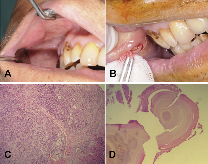
Results: Most of the patients were male (n = 14; 82.35%), with only three female patients (17.65%). The mean age of the 17 patients was 62.93 years (range, 35-82 years). Fifteen cases (∼88%) were found within the 6th-9th decades. Seven cases (∼41%) were identified in patients aged ≥70 years, six of which had been diagnosed in the most recent 5 years (2011-2015). The most common site was the buccal mucosa (n = 7; 41.18%), followed by the upper lip (n = 5; 29.41%), lower lip (n = 3; 17.65%), and vestibule and retromolar area (each n = 1; 5.88%). Only one case (5.88%) was clinically diagnosed as sialolithiasis prior to biopsy examination.
Conclusion: The current study demonstrated an aging tendency and a male predilection of sialolithiasis in minor salivary glands in Taiwan when compared with published case series studies.
Keywords: minor salivary gland; sialolith; sialolithiasis.
Figures
References
-
- Papin D. Salivary calculus. Dent Cosmos. 1864–5;6:136.
-
- Holst E. Sialolithiasis of the minor salivary glands: report of three cases. J Oral Surg. 1968;26:354–356. - PubMed
-
- Crawford W.H., Jr., Guernsey L.H. Sialolithiasis of minor salivary glands: report of case. J Oral Surg. 1969;27:649–652. - PubMed
-
- Hobkirk J.A. Sialolithiasis of a minor salivary gland. A case report and review of the literature. Dent Pract Dent Rec. 1970;20:213–214. - PubMed
-
- van der Waal I. Sialolithiasis of minor salivary glands: how rare? Report of two cases. J Oral Surg. 1971;29:815–816. - PubMed
LinkOut - more resources
Full Text Sources
