

Langerhans cells in 60 odontogenic keratocysts
- PMID: 30895063
- PMCID: PMC6399999
- DOI: 10.1016/j.jds.2017.04.001
Langerhans cells in 60 odontogenic keratocysts
Abstract
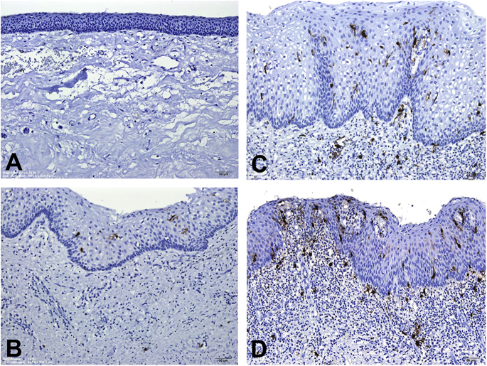
Background/purpose: Langerhans cells (LCs) are antigen-presenting cells. This study mainly evaluated the LC counts in 60 odontogenic keratocysts (OKCs).
Materials and methods: The CD1a-positive LC numbers in the lining epithelia and subepithelial connective tissues were counted at 60 OKC sites without inflammation, 39 OKC sites with mild/moderate inflammation, and 13 OKC sites with severe inflammation from 60 OKC specimens.
Results: The mean CD1a-positive LC counts in the lining epithelia and subepithelial connective tissues increased significantly from no inflammation (0.5 ± 0.4 and 0.2 ± 0.3 cell/high-power field or HPF, respectively) through mild/moderate inflammation (5.3 ± 2.5 and 2.5 ± 2.7 cells/HPF, respectively) to severe inflammation OKC sites (12.7 ± 5.6 and 9.3 ± 7.2 cells/HPF, respectively; all P-values < 0.001). OKC sites with inflammation had thicker lining epithelia than those without inflammation. Moreover, the mean CD1a-positive LC counts in the lining epithelia and subepithelial connective tissues of OKCs were significantly higher in the thicker lining epithelium (>100 μm) group (6.8 ± 5.1 and 3.7 ± 4.9 cells/HPF, respectively) than in the thinner lining epithelium (≦100 μm) group (1.0 ± 1.7 and 0.8 ± 2.5 cell/HPF, respectively; both P-values < 0.001).
Conclusion: There is a significant association of inflammation grade with the number of LCs in OKCs. The scarce LCs in the lining epithelia of OKCs without inflammation suggests the loss of immunosurveillance ability against the OKC lining epithelial cells; this can explain why OKCs have aggressive clinical behavior, a great growth potential, and a high recurrence rate.
Keywords: CD1a; Langerhans cell; aggressive clinical behavior; high recurrence rate; immunosurveillance; odontogenic keratocyst.
Figures
References
-
- Neville B.W., Damm D.D., Allen C.M., Chi A.C. Odontogenic cysts and tumors. In: Neville B.W., Damm D.D., Allen C.M., Chi A.C., editors. Oral and maxillofacial pathology. 4th ed. Elsevier; St. Louis: 2016. pp. 636–644.
-
- Neville B.W., Damm D.D., Allen C.M., Bouquot J.E. Odontogenic cysts and tumors. In: Neville B.W., Damm D.D., Allen C.M., Bouquot J.E., editors. Oral and maxillofacial pathology. 3rd ed. Saunders Elsevier; St. Louis: 2009. pp. 683–691.
-
- Philipsen H.P. Keratocystic odontogenic tumour. In: Barnes L., Eveson J.W., Reichart P., Sidransky D., editors. World Health Organization classification of tumours. Pathology and genetics of head and neck tumours. 3rd ed. IARC Press; Lyon: 2005. pp. 306–307.
-
- Speight P., Devilliers P., Li T.J., Odell E.W., Wright J.M. Odontogenic keratocyst. In: El-Naggar A.K., Chan J.K.C., Grandis J.R., Takata T., Slootweg P.J., editors. WHO classification of head and Neck tumours. 4th ed. IARC; Lyon: 2017. pp. 235–236.
-
- Barrett A.W., Cruchley A.T., Williams D.M. Oral mucosal Langerhans' cells. Crit Rev Oral Biol Med. 1996;7:36–58. - PubMed
LinkOut - more resources
Full Text Sources
