

doi: 10.1016/j.jds.2013.02.031.
Epub 2013 Sep 20.
Actinomycosis osteomyelitis of the jaws: Report of four cases and a review of the literature
Affiliations
- PMID: 30895066
- PMCID: PMC6400081
- DOI: 10.1016/j.jds.2013.02.031
Item in Clipboard
Actinomycosis osteomyelitis of the jaws: Report of four cases and a review of the literature
J Dent Sci.
2017 Sep.
Abstract
Actinomycosis osteomyelitis of the jaw bones, particularly in the maxilla, is an extremely rare disease. This report presents two cases of maxillary and two cases of mandibular actinomycosis osteomyelitis, with the diagnosis particularly based on histological procedures. The highly diversified pathogenicity of the phenomenon and the absence of solid diagnostic criteria are discussed. Laboratory challenges are emphasized, and a comprehensive overview of the entity including treatment alternatives is given along with a review of the relevant literature.
Keywords: actinomycosis; alveolar bone loss; cervicofacial; osteomyelitis.
Figures
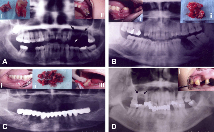
(A) Preoperative panoramic radiograph showing an extensive radiolucent lesion in the left maxillary premolar region (arrows). (i) Excised sequester and extracted neighboring molar tooth. (ii) Clinical view showing sequester and erythema of the gingiva in the maxillary premolar region. (B) Preoperative panoramic radiograph with radiolucent and sclerotic areas of the lesion in the left mandibular molar region. (i) Clinical view of an unhealed alveolar socket. (ii) Excisional biopsy material consisting of soft tissue and sequester. (C) Preoperative panoramic view showing radiolucent and radiopaque areas in the right mandibular premolar region below the prosthetic restoration. (i and iii) Preoperative clinical views showing gingival erythema and swelling of the right mandibular premolar region. (ii) Biopsy material. (D) Preoperative panoramic X-ray showing the right maxillary molar area with osteolytic and sclerotic lesion (arrows). (i) Clinical view of unhealed tooth socket and gingival erythema.
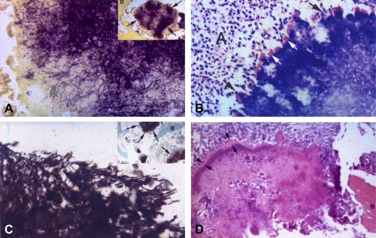
(A) Photomicrograph of peripherally radiating filaments of the actinomycotic granule (i), (tissue Gram stain, 100×). (ii) Granule is surrounded by trabecular bone. Note the deep-purple Gram-positive filaments (arrows), (tissue Gram stain, 20×). (B) Photomicrograph of deeply stained basophilic actinomycotic granule with prominent peripheral cubs (white arrows) surrounded by polymorphonuclear leucocytes (black arrows) embedded in an abscess (A), (Giemsa stain, 40×). (C) Periphery of actinomycotic granule (i). Note the branching filaments and cocoid elements. (ii) Gomori methamine silver stain of actinomycotic granule (arrows) embedded in an area of suppurative necrosis between bone trabeculae (10×). (D) Photomicrograph of an actinomycotic granule bordered by eosinophilic Splendore–Hoeppli material (between arrows), embedded in fibrino-purulent exudate and surrounded by trabecular bone (B). Note the numerous polymorphonuclear leucocytes within the matrix of the granule (H&E stain, 20×).
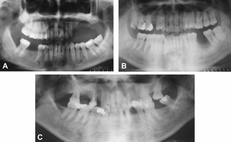
Postoperative panoramic radiographs showing the healing of bone in the (A) left maxillary premolar, (B) left mandibular molar, and (C) maxillary molar regions of Case 1, Case 2, and Case 4.
References
-
- Murray R.P., Kobayashi G.S., Pfaller M.A., Rosental K.S. 2nd ed. Mosby; St. Louis, MO: 1994. Medical Microbiology.
-
- Shafer W.G., Hine M.K., Levy B.M. 3rd ed. Saunders; Philadelphia, PA: 1974. A Textbook of Oral Pathology.
-
- Schuster G.S. 3rd ed. Decker; Philadelphia, PA: 1990. Oral Microbiology and Infectious Disease.
-
- Bennhoff D.F. Actinomycosis: diagnostic and therapeutic considerations and a review of 32 cases. Laryngoscope. 1984;94:1198–1217. - PubMed
-
- Ramachandran Nair P.N., Schroeder H.E. Periapical actinomycosis. J Endod. 1984;10:567–570. - PubMed
LinkOut - more resources
Full Text Sources
