

S100 protein-positive Langerhans cells in 80 dentigerous cysts
- PMID: 30895082
- PMCID: PMC6395349
- DOI: 10.1016/j.jds.2017.08.001
S100 protein-positive Langerhans cells in 80 dentigerous cysts
Abstract
Background/purpose: Langerhans cells (LCs) are antigen-presenting cells. This study assessed the LC counts in 80 dentigerous cysts (DCs).
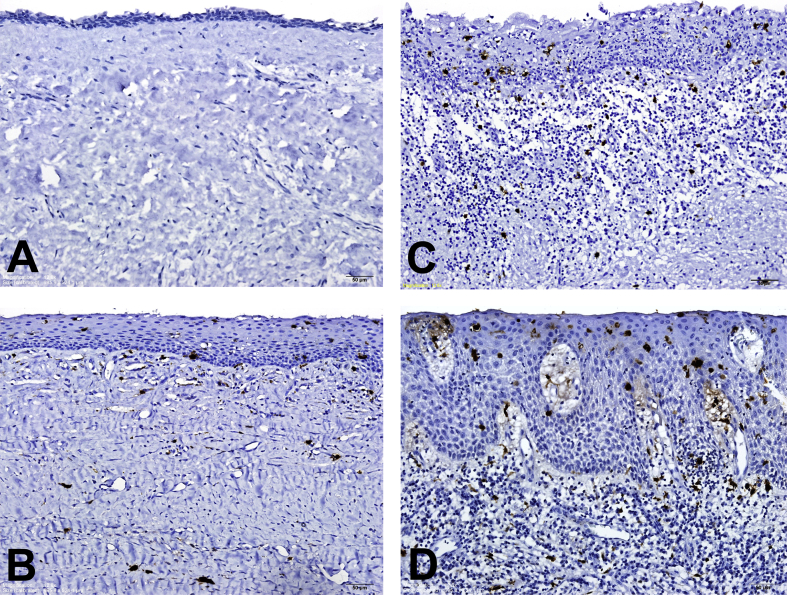
Materials and methods: The S100-positive LC numbers in the lining epithelia and subepithelial connective tissues were counted at 80 DC sites without inflammation, 33 DC sites with mild/moderate inflammation, and 9 DC sites with severe inflammation from 80 DC specimens.
Results: The mean S100-positive LC counts in the lining epithelia and subepithelial connective tissues increased significantly from no inflammation (0.6 ± 0.6 and 0.7 ± 0.6 cell/high-power field or HPF, respectively) through mild/moderate inflammation (8.1 ± 2.0 and 4.5 ± 2.3 cells/HPF, respectively) to severe inflammation DC sites (21.0 ± 7.0 and 11.1 ± 6.5 cells/HPF, respectively; P-value < 0.001). DC sites with inflammation had thicker lining epithelia than those without inflammation. Moreover, the mean LC counts in the lining epithelia and subepithelial connective tissues of DCs were significantly higher in the thicker lining epithelium (>50 μm) group (8.6 ± 7.1 and 4.8 ± 4.5 cells/HPF, respectively) than in the thinner lining epithelium (≦50 μm) group (0.6 ± 0.6 and 0.6 ± 0.6 cells/HPF, respectively; both P-values < 0.001).
Conclusion: A significant association of high-grade inflammation and thick lining epithelium with the increased LC number in DCs is found. Very few LCs in the lining epithelia of DCs without inflammation indicate the reduced immunosurveillance ability against DC lining epithelial cells in DC patients. It needs further studies to confirm the role of reduced immunosurveillance in the enlargement of the DC.
Keywords: Langerhans cell; dentigerous cyst; immunosurveillance; inflammation; lining epithelium.
Figures
References
-
- Neville B.W., Damm D.D., Allen C.M., Chi A.C. Odontogenic cysts and tumors. In: Neville B.W., Damm D.D., Allen C.M., Chi A.C., editors. Oral and Maxillofacial Pathology. 4th ed. Elsevier; St. Louis: 2016. pp. 632–635.
-
- Barrett A.W., Cruchley A.T., Williams D.M. Oral mucosal Langerhans' cells. Crit Rev Oral Biol Med. 1996;7:36–58. - PubMed
-
- Katz S.I., Tamaki K., Sachs D.H. Epidermal Langerhans cells are derived from cells originating in bone marrow. Nature. 1979;282:324–326. - PubMed
-
- Piattelli A., Rubini C., Iezzi G., Fioroni M. CD1a-positive cells in odontogenic cysts. J Endod. 2002;28:267–268. - PubMed
-
- Murase N., Tatemoto Y., Iwai Y., Okada Y., Mori M. Langerhans cells in odontogenic tumours and cysts as detected by S-100 protein immunohistochemistry. Basic Appl Histochem. 1990;34:135–141. - PubMed
LinkOut - more resources
Full Text Sources
