Review
. 2019 Mar 6:9:118.
doi: 10.3389/fonc.2019.00118. eCollection 2019.

Glioblastoma Stem- Like Cells, Metabolic Strategy to Kill a Challenging Target

Affiliations
Review

Glioblastoma Stem- Like Cells, Metabolic Strategy to Kill a Challenging Target

Delphine Garnier et al. Front Oncol. .

Abstract

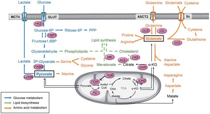
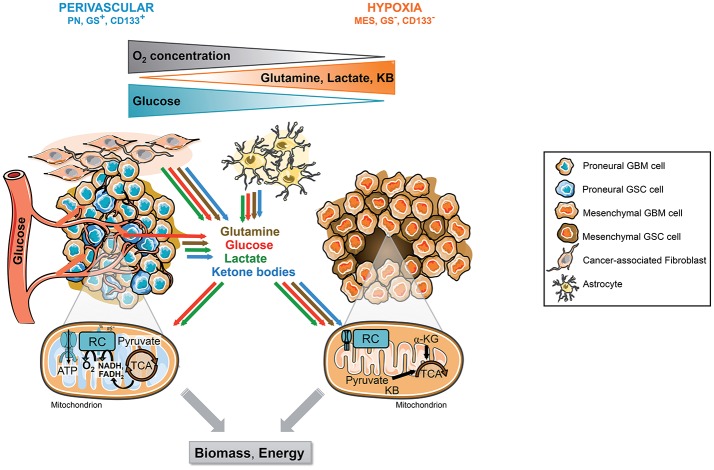
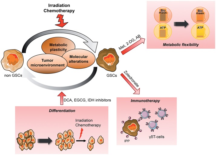
Over the years, substantial evidence has definitively confirmed the existence of cancer stem-like cells within tumors such as Glioblastoma (GBM). The importance of Glioblastoma stem-like cells (GSCs) in tumor progression and relapse clearly highlights that cancer eradication requires killing of GSCs that are intrinsically resistant to conventional therapies as well as eradication of the non-GSCs cells since GSCs emergence relies on a dynamic process. The past decade of research highlights that metabolism is a significant player in tumor progression and actually might orchestrate it. The growing interest in cancer metabolism reprogrammation can lead to innovative approaches exploiting metabolic vulnerabilities of cancer cells. These approaches are challenging since they require overcoming the compensatory and adaptive responses of GSCs. In this review, we will summarize the current knowledge on GSCs with a particular focus on their metabolic complexity. We will also discuss potential approaches targeting GSCs metabolism to potentially improve clinical care.

Keywords: Glioblastoma; cancer heterogeneity; cancer metabolism; cancer plasticity; cancer stem cells; tumor microenvironment.


Figures

Figure 1
Figure 1
Pyruvate and glutamate are 2 major metabolic hubs in GSCs. Tumor cells usually display a strong glycolytic metabolism. Glucose is uptaken by glucose transporter GLUT and then converted to pyruvate through several enzymes. All along this pathway, the glycolytic products are diverted from this main metabolic road to fuel other biosynthetic pathways such as the PPP as well as lipids and amino acids biosynthesis. PKM2 plays a key role in this dynamic process through conformational modulation. Glycolytic pyruvate will then either be converted to lactate or fuel mitochondrial OXPHOS and the TCA cycle. Another key metabolite that can fuel the TCA is glutamate once converted to αKG. Glutamate is produced either by GLS from glutamine or from glucose. Glutamate is involved in several biosynthetic pathways including amino acids and lipids biosynthesis as well as mitochondrial anaplerosis. Glutamate is also involved in glutathione synthesis, directly and indirectly by providing cysteine to the cells. α-KG, α-ketoglutarate; FAS, Fatty acid synthase; FBP1, Fructose-1,6 bisphosphatase 1; GCL, glutamate-cysteine ligase; GLS, Glutaminase; GS, Glutamine synthetase; GLUT, Glucose transporter 1; HK2, Hexokinase 2; IDH, Isocitrate dehydrogenase; LDHA, Lactate dehydrogenase A; MCT, Monocarboxylase transporter; MK, Mevalonate kinase; OAA, Oxaloacetate; PC, Pyruvate carboxylase; PDH, Pyruvate dehydrogenase; PDK, Pyruvate dehydrogenase Kinase; PKM2, Pyruvate kinase M2; PPP, Pentose phosphate pathway; TCA, Tricarboxylic acid cycle.
Figure 2
Figure 2
Tumor microenvironment and molecular signature, 2 drivers of GSCs metabolic phenotype. GSCs reside in singular tumor microenvironment in GBM called the perivascular and the hypoxic niche. Each niche is associated with specific molecular signature and metabolic phenotype. The perivascular GSCs display a PN signature and a strong glycolytic metabolism mainly based on blood glucose. In these cells, glucose can be directly converted to lactate or oxidized within the mitochondria through OXPHOS. Furthermore, these cells highly express GS allowing the direct synthesis and secretion of glutamine within the TME. In contrast, hypoxic GSCs usually belong to the mesenchymal subtype and do not express GS. These cells are residing in a harsher environment display flexible metabolism fueled by lactate, ketone bodies, amino acids, including glutamine, released in the microenvironment by other cancer cells and/or stromal cells including astrocytes or cancer-associated fibroblasts. In absence of O2, these substrates supply a truncated TCA cycle allowing the generation of energy and biosynthetic precursors such as lipids or nucleic acids, as well as antioxidant glutathione synthesis. CAF, Cancer-associated fibroblast; GBM, Glioblastoma; GSCs, Glioblastoma stem-like cells, GS, Glutamine synthetase; αKG, Alpha-ketoglutarate; KB, Ketone bodies; MES, Mesenchymal; OXPHOS, Mitochondrial oxidative phosphorylation; PN, Proneural; TCA, Tricarboxylic acid cycle.
Figure 3
Figure 3
Metabolic targeting of the dynamic emergence of GSCs from differentiated GBM GSCs emerge from differentiated tumor cells, which can reacquire stem cells properties such as self-renewal, multilineage differentiation and the ability to give rise to the initial heterogenous tumor in vivo. This dynamic differentiation/dedifferentiation balance is driven by the TME, molecular events as well as their metabolic phenotype. Thus, metabolic targeted therapy appears as a potential novel therapeutic avenue, in particular when combined to conventional treatment such as radiation therapy. Several angles can be taken to eradicate GSCs from the tumor, reducing their metabolic plasticity and/or metabolic cooperation with the TME, inducing their differentiation to sensitize them to treatment or increasing their recognition by immune effectors. AB, Antibiotics; CAF, Cancer-associated fibroblast; DCA, Dichloroacetate; 2-DG, 2-Deoxyglucose; EGCG, Epigallocatechine gallate; GSCs, Glioblastoma Stem-like cells; IPP, Isopentenyl pyrophosphate; Met, Metformin; MSC, Mesenchymal stem cell.

References

    1. Louis DN, Perry A, Reifenberger G, von Deimling A, Figarella-Branger D, Cavenee WK, et al. . The 2016 World Health Organization classification of tumors of the central nervous system: a summary. Acta Neuropathol. (2016) 131:803–20. 10.1007/s00401-016-1545-1 - DOI - PubMed
    1. Stupp R, Hegi ME, Mason WP, van den Bent MJ, Taphoorn MJ, Janzer RC, et al. . Effects of radiotherapy with concomitant and adjuvant temozolomide versus radiotherapy alone on survival in glioblastoma in a randomised phase III study: 5-year analysis of the EORTC-NCIC trial. Lancet Oncol. (2009) 10:459–66. 10.1016/S1470-2045(09)70025-7 - DOI - PubMed
    1. Gusyatiner O, Hegi ME. Glioma epigenetics: from subclassification to novel treatment options. Semin Cancer Biol. (2018) 51:50–8. 10.1016/j.semcancer.2017.11.010 - DOI - PubMed
    1. Garnier D, Meehan B, Kislinger T, Daniel P, Sinha A, Abdulkarim B, et al. . Divergent evolution of temozolomide resistance in glioblastoma stem cells is reflected in extracellular vesicles and coupled with radiosensitization. Neuro Oncol. (2018) 20:236–48. 10.1093/neuonc/nox142 - DOI - PMC - PubMed
    1. Johnson BE, Mazor T, Hong C, Barnes M, Aihara K, McLean CY, et al. . Mutational analysis reveals the origin and therapy-driven evolution of recurrent glioma. Science. (2014) 343:189–93. 10.1126/science.1239947 - DOI - PMC - PubMed