. 2019 May;32(3):492-503.
doi: 10.1007/s10548-019-00703-1. Epub 2019 Mar 20.

MEG Assessment of Expressive Language in Children Evaluated for Epilepsy Surgery

Affiliations

MEG Assessment of Expressive Language in Children Evaluated for Epilepsy Surgery

Elaine Foley et al. Brain Topogr. 2019 May.

Abstract

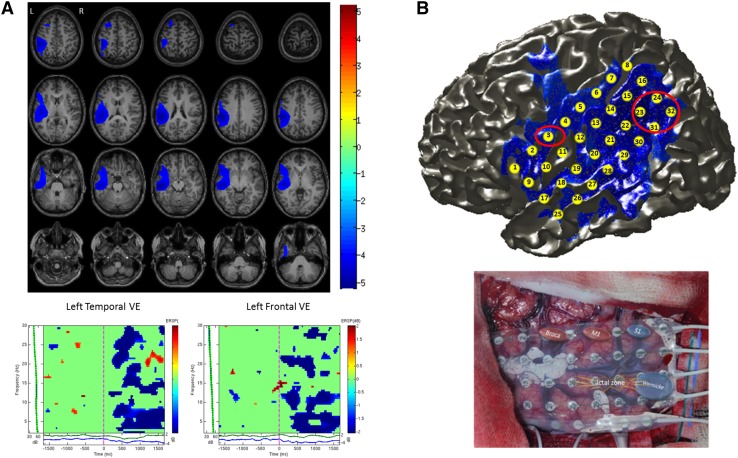
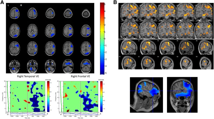
Establishing language dominance is an important step in the presurgical evaluation of patients with refractory epilepsy. In the absence of a universally accepted gold-standard non-invasive method to determine language dominance in the preoperative assessment, a range of tools and methodologies have recently received attention. When applied to pediatric age, many of the proposed methods, such as functional magnetic resonance imaging (fMRI), may present some challenges due to the time-varying effects of epileptogenic lesions and of on-going seizures on maturational phenomena. Magnetoencephalography (MEG) has the advantage of being insensitive to the distortive effects of anatomical lesions on brain microvasculature and to differences in the metabolism or vascularization of the developing brain and also provides a less intimidating recording environment for younger children. In this study we investigated the reliability of lateralized synchronous cortical activation during a verb generation task in a group of 28 children (10 males and 18 females, mean age 12 years) with refractory epilepsy who were evaluated for epilepsy surgery. The verb generation task was associated with significant decreases in beta oscillatory power (13-30 Hz) in frontal and temporal lobes. The MEG data were compared with other available presurgical non-invasive data including cortical stimulation, neuropsychological and fMRI data on language lateralization where available. We found that the lateralization of MEG beta power reduction was concordant with language dominance determined by one or more different assessment methods (i.e. cortical stimulation mapping, neuropsychological, fMRI or post-operative data) in 89% of patients. Our data suggest that qualitative hemispheric differences in task-related changes of spectral power could offer a promising insight into the contribution of dominant and non-dominant hemispheres in language processing and may help to characterize the specialization and lateralization of language processes in children.

Keywords: Beamformers; Children; Epilepsy surgery; Functional mapping; Hemispheric dominance; Language lateralization; Magnetoencephalography.


Figures

Fig. 1
Fig. 1
a Beamformer source localization results of verb generation task for an individual patient (Pt. 13) are shown overlaid on axial slices of the patient’s T1 MRI. Sources showing significant decreases in beta oscillatory power (13–30 Hz) from 2.5 s pre- to 2.5 s post-stimulus onset were identified in left temporal and left frontal regions (LI = 64.95). Time–frequency analysis of a virtual electrode (VE) constructed in the left temporal lobe shows an early sustained decrease in beta power during covert verb generation. Similarly, time–frequency analysis of a virtual electrode constructed in the left frontal lobe shows decreases in beta power from around 200 ms during covert verb generation. b Top: Electrode grid co-registered and overlaid on cortical surface of the patients MRI along with MEG beamformer source localization results. Red circles indicate location of Wernicke’s and Broca’s areas as identified during direct cortical stimulation. The peak source of decreased beta power (13–30 Hz) localized by MEG corresponds to Wernicke’s area in this patient. Bottom: Photograph of 32-channel grid shows results from cortical stimulation, where Broca’s area was mapped to contact 3 and Wernicke’s area to contacts 23–24 and 31–32. When these contacts were stimulated the patient had difficulty producing verbs, was confused and stuttered
Fig. 2
Fig. 2
a Beamformer source localization results of verb generation task for an individual patient (Pt. 11) are shown overlaid on axial slices of the patient’s T1 MRI. Sources showing significant decreases in beta oscillatory power (13–30 Hz) from 2.5 s pre- to 2.5 s post-stimulus onset were identified in right temporal and right frontal regions (LI = − 47.91). Time–frequency analysis of a virtual electrode (VE) constructed in the right temporal lobe shows significant decreases in beta power from approximately 300 ms post-stimulus onset. Similarly, time–frequency analysis of a virtual electrode constructed in the right frontal lobe shows significant decreases in beta power from approximately 300 ms post-stimulus onset. b Sources of decreased beta power identified with MEG correspond with fMRI activation during the same covert verb generation task showing right lateralized activation. fMRI activation is shown overlaid on coronal and sagittal slices of the patients MRI. Blue cross-hairs indicate region of peak decrease in MEG beta power in the right inferior frontal gyrus which corresponds to area of increased activation in the fMRI data

Similar articles

Cited by

References

    1. Abou-Khalil B. An update on determination of language dominance in screening for epilepsy surgery: the Wada test and newer. noninvasive alternatives. Epilepsia. 2007;48:442–455. doi: 10.1111/j.1528-1167.2007.01012.x. - DOI - PubMed
    1. Adjamian P, et al. Co-registration of magnetoencephalography with magnetic resonance imaging using bite-bar-based fiducials and surface-matching. Clin Neurophysiol. 2004;115:691–698. doi: 10.1016/j.clinph.2003.10.023. - DOI - PubMed
    1. Bagic AI, Bowyer SM, Kirsch HE, Funke ME, Burgess RC, Committee APS. American Clinical MEG Society (ACMEGS) Position Statement #2: The Value of Magnetoencephalography (MEG)/Magnetic Source Imaging (MSI) in noninvasive presurgical mapping of eloquent cortices of patients preparing for surgical interventions. J Clin Neurophysiol. 2017;34:189–195. doi: 10.1097/WNP.0000000000000366. - DOI - PubMed
    1. Balter S, Lin G, Leyden KM, Paul BM, McDonald CR. Neuroimaging correlates of language network impairment and reorganization in temporal lobe epilepsy. Brain Lang. 2016 - PMC - PubMed
    1. Berl MM, et al. Characterization of atypical language activation patterns in focal epilepsy. Ann Neurol. 2014;75:33–42. doi: 10.1002/ana.24015. - DOI - PMC - PubMed

Publication types

LinkOut - more resources