. 2019 Oct 1;145(7):1921-1934.
doi: 10.1002/ijc.32293. Epub 2019 Apr 4.

Nuclear orphan receptor NR2F6 confers cisplatin resistance in epithelial ovarian cancer cells by activating the Notch3 signaling pathway

Affiliations

Nuclear orphan receptor NR2F6 confers cisplatin resistance in epithelial ovarian cancer cells by activating the Notch3 signaling pathway

Han Li et al. Int J Cancer. .

Abstract

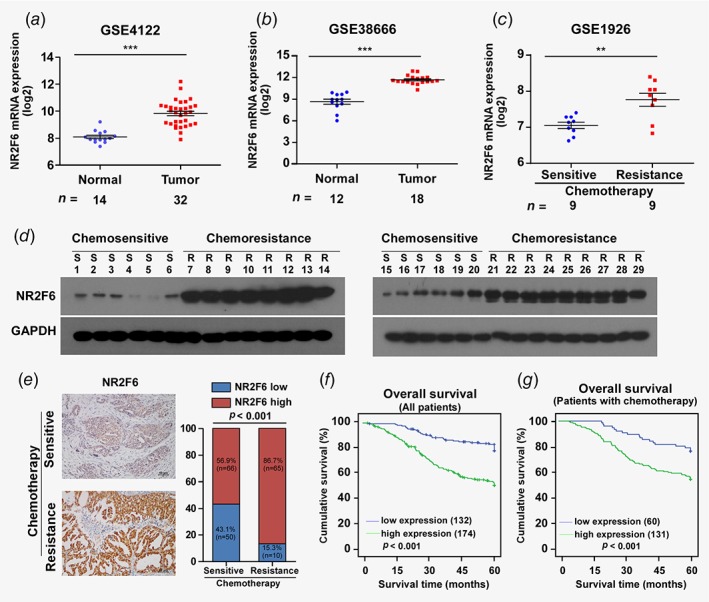
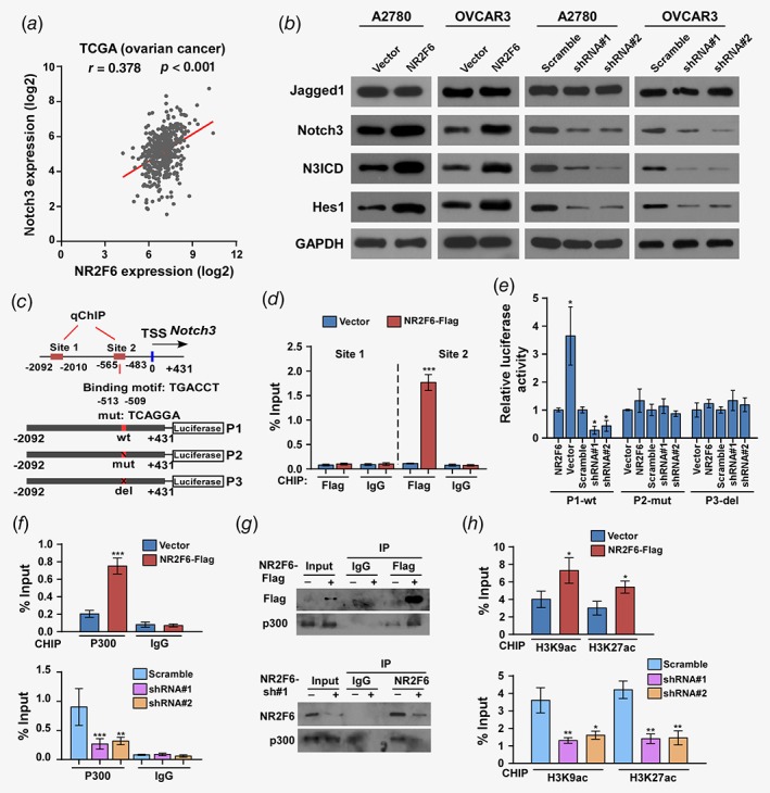
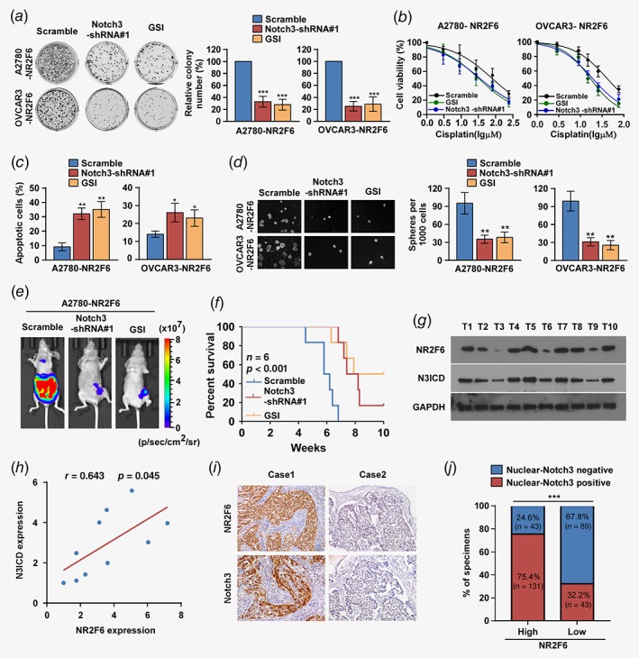
The primary challenge facing treatment of epithelial ovarian cancer (EOC) is the high frequency of chemoresistance, which severely impairs the quality of life and survival of patients with EOC. Our study aims to investigate the mechanisms by which upregulation of NR2F6 induces chemoresistance in EOC. The biological roles of NR2F6 in EOC chemoresistance were explored in vitro by Sphere, MTT and AnnexinV/PI assay, and in vivo using an ovarian cancer orthotopic transplantation model. Bioinformatics analysis, luciferase assay, CHIP and IP assays were performed to identify the mechanisms by which NR2F6 promotes chemoresistance in EOC. The expression of NR2F6 was significantly upregulated in chemoresistant EOC tissue, and NR2F6 expression was correlated with poorer overall survival. Moreover, overexpression of NR2F6 promotes the EOC cancer stem cell phenotype; conversely, knockdown of NR2F6 represses the EOC cancer stem cell phenotype and sensitizes EOC to cisplatin in vitro and in vivo. Our results further demonstrate that NR2F6 sustains activated Notch3 signaling, resulting in chemoresistance in EOC cells. Notably, NR2F6 acts as an informative biomarker to identify the population of EOC patients who are likely to experience a favorable objective response to gamma-secretase inhibitors (GSI), which inhibit Notch signaling. Therefore, concurrent inhibition of NR2F6 and treatment with GSI and cisplatin-based chemotherapy may be a novel therapeutic approach for NR2F6-overexpressing EOC. In summary, we have, for the first time, identified an important role for NR2F6 in EOC cisplatin resistance. Our study suggests that GSI may serve as a potential targeted treatment for patients with NR2F6-overexpressing EOC.

Keywords: NR2F6; Notch3 signaling pathway; cancer stem cells; chemoresistance; epithelial ovarian cancer.


Figures

Figure 1
Figure 1
NR2F6 is overexpressed in EOC and correlates with chemoresistance. (a) Expression profiling of mRNAs in EOC tumor vs. normal tissue samples and chemosensitive tissues vs. chemoresistant tissues from Array express microarray data (GEO accession number: GSE4122, GSE38666 and GSE1926; http://www.ncbi.nlm.nih.gov/geo/). (b,c) Western blot analysis of NR2F6 protein in 29 primary ovarian cancer tissues, including 12 samples that had no chemoresistance observed within 6 months, and 17 samples with chemoresistance within 6 months. GAPDH was used as a loading control. Epithelial ovarian cancer (EOC) patients whose disease recurs in less than 6 months after initial platinum‐based chemotherapy are termed platinum resistance, and the recurrence of EOC patients after 6 months is considered platinum sensitive (Ovarian Cancer National Comprehensive Cancer Network (NCCN) 2017). (d) Representative images of EOC tumor samples with (n = 75) or without (n = 116) chemoresistance in 6 months (left panel). Correlation between chemoresistance and NR2F6 expression in patients; the Chi‐square test was used to evaluate correlation (right panel). *p < 0.05,**p < 0.01, ***p < 0.001. [Color figure can be viewed at wileyonlinelibrary.com]
Figure 2
Figure 2
Overexpression of NR2F6 confers cisplatin resistance to ovarian cancer in vitro. (a) Representative images (left panel) and quantification (right panel) of the indicated cells after crystal violet staining. Each bar represents the mean ± SD of three independent experiments. *p < 0.05; **p < 0.01. (b) MTT/IC50 of cisplatin in the indicated cells. Each bar represents the mean ± SD of three independent experiments. *p < 0.05. (c) Annexin V‐FITC and PI staining of the indicated cells treated with cisplatin for 24 hr. Each bar represents the mean ± SD of three independent experiments. *p < 0.05,**p < 0.01. Abbreviations: FITC, fluorescein isothiocyanate; PI, propidium iodide. (d) Western blot analysis of cleaved caspase3 and PARP in the indicated cells; GAPDH was used as a loading control. [Color figure can be viewed at wileyonlinelibrary.com]
Figure 3
Figure 3
Overexpression of NR2F6 confers cisplatin resistance on ovarian cancer in vivo. (a) Representative images of tumor‐bearing mice challenged with the indicated cells treated with 5 mg/kg cisplatin. (b) Relative changes in luminescence were evaluated by bioluminescent imaging (BLI). (c) Kaplan–Meier survival curves of mice in the indicated cell. (df) HE and IHC staining were used in indicated xenografts. Each bar represents the mean ± SD of three independent experiments. *p < 0.05,**p < 0.01. [Color figure can be viewed at wileyonlinelibrary.com]
Figure 4
Figure 4
Overexpression of NR2F6 confers cancer stem cell characteristics on EOC cells in vitro and in vivo. (a) Representative images of tumor spheres formed by the indicated A2780 and OVCAR3 cells. (b) Histograms showing the mean number of spheres formed by the indicated cells from different passages. (c) Fold change in the number of cells per sphere on the indicated days. *p < 0.05, **p < 0.01, ***p < 0.001. (d) Statistical analysis of tumor incidence and tumor‐initiating cell (TIC) frequency calculated by ELDA software. (e, f) Limiting dilution assays to analyze the effect of NR2F6 on ovarian cancer tumorigenesis. (e) Indicated numbers of A2780 cells were implanted into the flank of BALB/c nude mice; tumor volumes were measured on the indicated days. (f) Five weeks later, mice were euthanized and tumors were retrieved and weighed. [Color figure can be viewed at wileyonlinelibrary.com]
Figure 5
Figure 5
NR2F6 regulates Notch3 transcriptional activity in ovarian cancer. (a) Correlation between NR2F6 and Notch3 mRNA expression in TCGA ovarian cancer dataset. (b) Expression of Jagged1, Notch3, N3ICD, and Hes1 in the indicated A2780 and OVCAR3 cells were evaluated by western blot. GAPDH was used as loading control. (c) Schematic illustration of the PCR fragments of the human Notch3 gene promoter. (d) Chromatin immunoprecipitation (ChIP) assays were performed in A2780 cells using antibodies against Flag to identify the NR2F6 occupancy on the Notch3 gene promoter. Immunoglobulin G (IgG) was used as a negative control. (e) Luciferase reporter assays were conducted in the indicated A2780 cells to detect the effect of deletion or mutation of site 2 fragment of the Notch3 promoters on transcriptional regulation by NR2F6. (f) ChIP assays were performed in the indicated A2780 cells using antibodies against p300 acetyltransferase to identify the p300 occupancy on the Notch3 gene promoters. Immunoglobulin G (IgG) was used as a negative control. (g) Lysates from A2780 cells transfected with Flag‐NR2F6 were immunoprecipitated with anti‐Flag affinity agarose, followed by western blot analysis of p300. Immunoprecipitation assays revealed that silencing of NR2F6 substantially abrogated the interaction with p300. (h) Enrichments acetylated histone H3 (K9, K27) in site 2 fragment of the Notch3 gene promoter in the indicated A2780 cells. *p < 0.05, **p < 0.01, ***p < 0.001. [Color figure can be viewed at wileyonlinelibrary.com]
Figure 6
Figure 6
Notch3 is required for NR2F6‐induced cisplatin resistance and the clinical relevance of NR2F6 and Notch3. (a) Representative images (left panel) and quantification (right panel) after crystal violet staining of the indicated cells treated with Notch3 shRNAs and Notch3 inhibitor (GSI). (b) MTT/IC50 of cisplatin in the indicated cells treated with Notch3 shRNAs and Notch3 inhibitor (GSI). Each bar represents the mean ± SD of three independent experiments. *p < 0.05. (c). Annexin V‐FITC and PI staining of the indicated cells treated with Notch3 shRNAs and Notch3 inhibitor (GSI). Each bar represents the mean ± SD of three independent experiments. (d) Representative micrographs and quantification of the tumor spheres formed by A2780‐NR2F6 and OVCAR3‐NR2F6 cells subjected to the indicated treatments. (e, f) Representative images (e) and Kaplan–Meier survival curves (f) of tumor‐bearing mice subjected to Notch3 shRNAs and Notch3 inhibitor (GSI) treatments. (g) Expression analysis and (h) correlation of NR2F6 and Notch3 expression in 10 freshly collected human ovarian cancer tissue samples; GAPDH was used as a loading control. Each bar represents the mean ± SD of three independent experiments. (i, j) Immunohistochemical staining showing that NR2F6 expression correlates positively with Notch3 expression in 306 clinical ovarian cancer specimens. Percentage of human ovarian cancer specimens showing low or high NR2F6 expression relative to the levels of Notch3. Scale bars = 50 μm; ***p < 0.001. [Color figure can be viewed at wileyonlinelibrary.com]

References

    1. Siegel RL, Miller KD, Jemal A. Cancer statistics, 2017. CA Cancer J Clin 2017;67:7–30. - PubMed
    1. Mezzanzanica D. Ovarian cancer: a molecularly insidious disease. Chin J Cancer 2015;34:1–3. - PMC - PubMed
    1. Nick AM, Coleman RL, Ramirez PT, et al. A framework for a personalized surgical approach to ovarian cancer. Nat Rev Clin Oncol 2015;12:239–45. - PMC - PubMed
    1. Amate P, Huchon C, Dessapt AL, et al. Ovarian cancer: sites of recurrence. Int J Gynecol Cancer 2013;23:1590–6. - PubMed
    1. Tsai SY, Tsai MJ. Chick ovalbumin upstream promoter‐transcription factors (COUP‐TFs): coming of age. Endocr Rev 1997;18:229–40. - PubMed

Publication types

MeSH terms