. 2019 Jun;10(3):630-642.
doi: 10.1002/jcsm.12409. Epub 2019 Mar 20.

Fibroblast growth factor 21 controls mitophagy and muscle mass

Affiliations

Fibroblast growth factor 21 controls mitophagy and muscle mass

Lynette J Oost et al. J Cachexia Sarcopenia Muscle. 2019 Jun.

Abstract

Background: Skeletal muscle is a plastic tissue that adapts to changes in exercise, nutrition, and stress by secreting myokines and myometabolites. These muscle-secreted factors have autocrine, paracrine, and endocrine effects, contributing to whole body homeostasis. Muscle dysfunction in aging sarcopenia, cancer cachexia, and diabetes is tightly correlated with the disruption of the physiological homeostasis at the whole body level. The expression levels of the myokine fibroblast growth factor 21 (FGF21) are very low in normal healthy muscles. However, fasting, ER stress, mitochondrial myopathies, and metabolic disorders induce its release from muscles. Although our understanding of the systemic effects of muscle-derived FGF21 is exponentially increasing, the direct contribution of FGF21 to muscle function has not been investigated yet.

Methods: Muscle-specific FGF21 knockout mice were generated to investigate the consequences of FGF21 deletion concerning skeletal muscle mass and force. To identify the mechanisms underlying FGF21-dependent adaptations in skeletal muscle during starvation, the study was performed on muscles collected from both fed and fasted adult mice. In vivo overexpression of FGF21 was performed in skeletal muscle to assess whether FGF21 is sufficient per se to induce muscle atrophy.

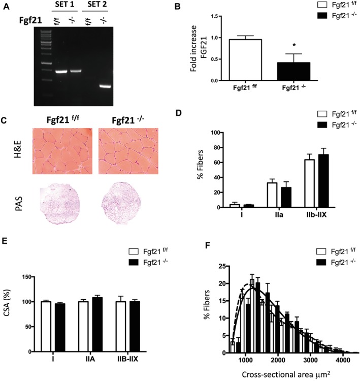
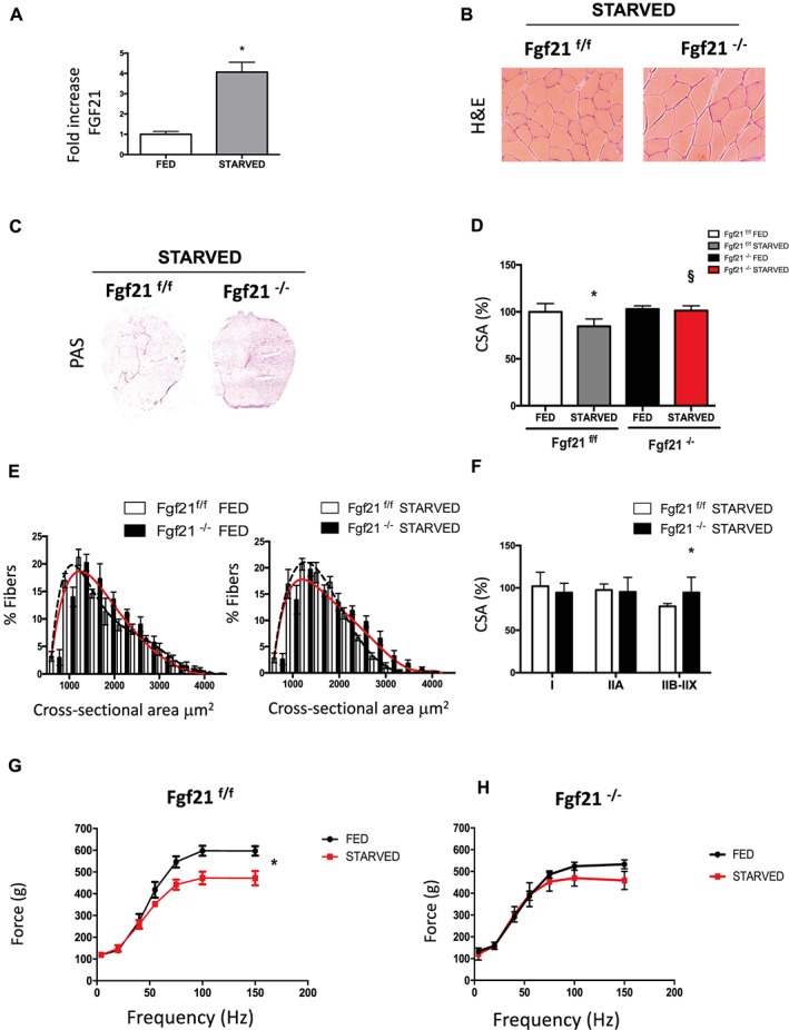
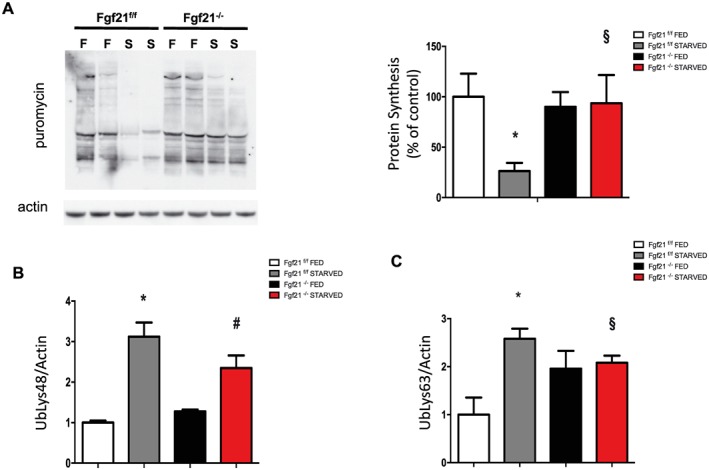
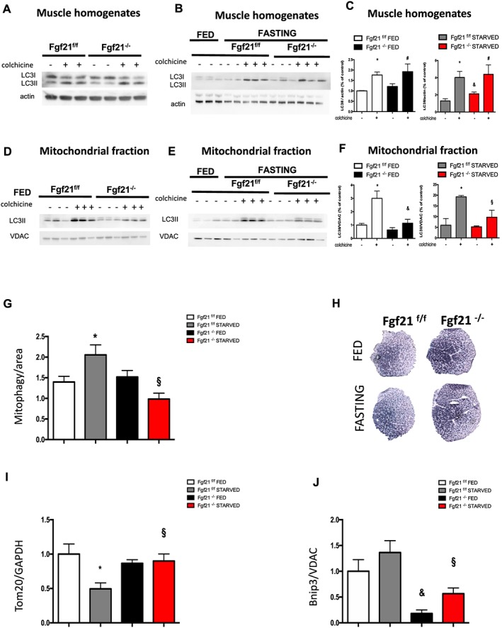
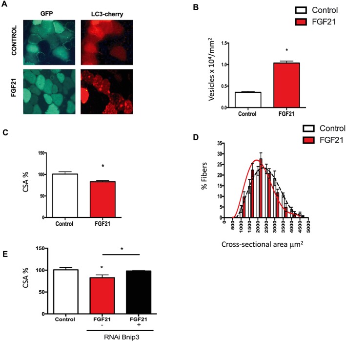
Results: We show that FGF21 does not contribute to muscle homeostasis in basal conditions in terms of fibre type distribution, fibre size, and muscle force. In contrast, FGF21 is required for fasting-induced muscle atrophy and weakness. The mass of isolated muscles from control-fasted mice was reduced by 15-25% (P < 0.05) compared with fed control mice. FGF21-null muscles, however, were significantly protected from muscle loss and weakness during fasting. Such important protection is due to the maintenance of protein synthesis rate in knockout muscles during fasting compared with a 70% reduction in control-fasted muscles (P < 0.01), together with a significant reduction of the mitophagy flux via the regulation of the mitochondrial protein Bnip3. The contribution of FGF21 to the atrophy programme was supported by in vivo FGF21 overexpression in muscles, which was sufficient to induce autophagy and muscle loss by 15% (P < 0.05). Bnip3 inhibition protected against FGF21-dependent muscle wasting in adult animals (P < 0.05).

Conclusions: FGF21 is a novel player in the regulation of muscle mass that requires the mitophagy protein Bnip3.

Keywords: Autophagy; Bnip3; FGF21; Mitophagy; Muscle atrophy; Myokine.


Conflict of interest statement

None declared.

Figures

Figure 1
Figure 1
Muscle‐specific FGF21 deletion does not affect muscle size or histology. (A) Genotyping of FGF21 control and knockout mice. PCR analysis with genomic DNA from gastrocnemius muscles. Two sets of primers were used. Set 1 consists of one primer upstream of the 5′ loxP site and the other primer inside FGF21 sequence, amplifying a 675 bp sequence corresponding to not deleted DNA; set 2 consists of a couple of primers upstream 5′ loxP and downstream 3′ loxP sites that detects the deleted sequence. (B) FGF21 mRNA expression was quantified by q‐PCR in tibialis anterior muscle of FGF21−/− and control mice. (C) Representative H&E and PAS staining showing normal morphology, and glycogen content of FGF21−/− gastrocnemius muscle. (D) Percentage of fibres expressing myosin heavy chain types I, IIA, IIB, and IIX proteins in gastrocnemius muscles revealed by immunohistochemistry analysis. (E) Quantification of CSA of myofibers indicates no significant differences in FGF21‐ablated muscles. (F) Frequency histograms showing the distribution of cross‐sectional areas (μm2) in GNM of FGF21f/f (black dashed line) and FGF21−/− (black line) fibres. Data are shown as mean ± SEM. * P < 0.05. CSA, cross‐sectional area; FGF21, fibroblast growth factor 21; H&E, haematoxylin and eosin; PAS, periodic acid–Schiff.
Figure 2
Figure 2
FGF21 is required for muscle atrophy and weakness during fasting. (A) mRNA expression of FGF21 in GNM of fed and starved FGF21f/f mice. (B) H&E staining of control (FGF21f/f) and KO mice (FGF21−/−) GNM muscle. (C) PAS staining shows more glycogen content in FGF21 null muscles. (D) Cross‐sectional area of fed and 48 h starved GNM muscles. (E) Fibre size distribution from fed (left panel) and starved (right panel) control (black dashed line) and FGF21 KO (red line) muscles. (F) CSA of myofibers expressing myosin heavy chain types IIA and IIB‐IIX of starved control and KO mice. (G) Force measurements performed in vivo on gastrocnemius showed that FGF21−/− muscles preserve muscle force after fasting. Force/frequency curve of FGF21f/f (left panel) and FGF21−/− (right panel). Data are shown as mean ± SEM. Significance P < 0.05 *vs. control fed, §vs. control starved. CSA, cross‐sectional area; FGF21, fibroblast growth factor 21; H&E, haematoxylin and eosin; KO, knockout; PAS, periodic acid–Schiff.
Figure 3
Figure 3
Protein synthesis rate is maintained in FGF21‐deleted muscles during fasting. (A) In vivo surface sensing of translation technique shows the maintenance of protein synthesis in FGF21‐ablated mice muscles during fasting. A representative immunoblot is shown. Quantification of the puromycin‐labelled peptides is expressed as percentage of the values obtained in the control group. Data are normalized to actin. (B–C) Densitometric analysis of total muscle extracts from FGF21f/f and FGF21−/−immunoblotted for anti‐ubiquitin (Lys48) (B) and for anti‐ubiquitin (Lys63) (C) normalized to actin. Data are shown as mean ± SEM. Protein bands were quantified using ImageJ, normalized to actin, and their expression was plotted relative to control fed (set to 1.00). Significance: *compared with control fed (P < 0.01), and compared with control fed (P < 0.05), #compared to FGF21 knockout fed (P < 0.05) and §vs. control starved (P < 0.05). FGF21, fibroblast growth factor 21.
Figure 4
Figure 4
Muscle‐specific FGF21 deletion controls mitophagy. Immunoblotting analyses of autophagy flux in control and FGF21‐null GNM total muscle homogenates in fed (A) and in fasting (B) conditions. (C) Quantification of LC3 lipidation was normalized to actin and plotted to control fed not treated with colchicine (set to 1.00). Inhibition of autophagy–lysosome fusion by colchicine treatment induces less accumulation of LC3II band in starved FGF21‐KO but not in starved control muscles. Mitophagy flux is decreased in FGF21 KO muscles. (D–E) Representative immunoblot images of mitochondria isolated from GNM muscles after colchicine treatment probed with anti‐LC3 antibody in fed (D) and in fasting conditions. (F) Quantification of protein bands as seen in (D and E) by ImageJ. VDAC served as a loading control. (G) Mitophagy flux was analysed by electroporation of a reporter plasmid (mt‐mKEIMA) into flexor digitorum brevis muscles of adult control and FGF21−/−mice; changes of fluorescent spectra were detected and normalized to fibre area. (H) Succinate dehydrogenase staining indicating more mitochondrial content in FGF21 KO GNM muscles in both fed and fasting. (I) Mitochondrial content revealed by Tom20 is increased in FGF21‐deleted muscles during fasting. Densitometric quantification of Tom20 western blot, normalized to GAPDH. (J) The mitophagy protein Bnip3 is significantly reduced in KO muscles. Densitometric analysis of Bnip3 protein, normalized to the mitochondrial protein VDAC and plotted relative to control fed. Data are shown as mean ± SEM. Significance P < 0.05 *compared with control fed and compared with control fed and §compared with control starved. FGF21, fibroblast growth factor 21; GNM, gastrocnemius; KO, knockout.
Figure 5
Figure 5
In vivo overexpression of FGF21 in muscles induces autophagy and Bnip3‐dependent muscle atrophy. (A–B) FGF21 overexpression induces autophagy. (A) Representative images of adult tibialis anterior muscles of wild type mice electroporated in vivo with mcherry‐LC3 and with either GFP‐FGF21 or with only GFP (control). Muscles were collected 12 days after transfection. (B) A higher number of LC3 dots was observed in muscles overexpressing FGF21. Quantification of the number of LC3 positive vesicles normalized to fibre area in control or in FGF21 overexpressing muscles. (C) FGF21 in vivo overexpression is sufficient to induce muscle loss. CSA of transfected fibres, identified by GFP immunofluorescence, was measured with ImageJ and normalized to control fibres. (D) Frequency histograms showing the distribution of cross‐sectional areas (μm2) in tibialis anterior transfected with either a GFP‐plasmid (control) (black dashed line) or with GFP‐FGF21 plasmid (red line). (E) Downregulation of Bnip3, by RNAi protects from FGF21‐dependent muscle loss. Adult skeletal muscles were co‐transfected with either GFP or with FGF21 in the presence or absence of specific siRNAs for mouse Bnip3. Twelve days later, muscles were collected and analysed for CSA of transfected fibres. Data are shown as mean ± SEM. * P < 0.05. CSA, cross‐sectional area; FGF21, fibroblast growth factor 21; GFP, green fluorescent protein.

Comment in

References

    1. Volaklis KA, Halle M, Meisinger C. Muscular strength as a strong predictor of mortality: a narrative review. Eur J Intern Med 2015;26:303–310. - PubMed
    1. Romanello V, Sandri M. Mitochondrial quality control and muscle mass maintenance. Front Physiol 2015;6:422. - PMC - PubMed
    1. Rai M, Demontis F. Systemic nutrient and stress signaling via myokines and myometabolites. Annu Rev Physiol 2016;78:85–107. - PubMed
    1. Whitham M, Febbraio MA. The ever‐expanding myokinome: discovery challenges and therapeutic implications. Nat Rev Drug Discov 2016;15:719–729. - PubMed
    1. Pedersen BK, Steensberg A, Fischer C, Keller C, Keller P, Plomgaard P, et al. Searching for the exercise factor: is IL‐6 a candidate? J Muscle Res Cell Motil 2003;24:113–119. - PubMed

Publication types

LinkOut - more resources