

Accuracy of deep learning to differentiate the histopathological grading of meningiomas on MR images: A preliminary study
- PMID: 30896065
- PMCID: PMC6767062
- DOI: 10.1002/jmri.26723
Accuracy of deep learning to differentiate the histopathological grading of meningiomas on MR images: A preliminary study
Abstract
Background: Grading of meningiomas is important in the choice of the most effective treatment for each patient.
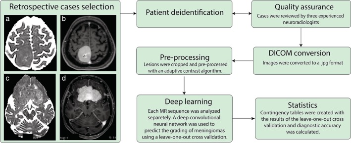
Purpose: To determine the diagnostic accuracy of a deep convolutional neural network (DCNN) in the differentiation of the histopathological grading of meningiomas from MR images.
Study type: Retrospective.
Population: In all, 117 meningioma-affected patients, 79 World Health Organization [WHO] Grade I, 32 WHO Grade II, and 6 WHO Grade III.
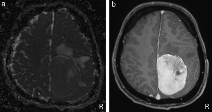
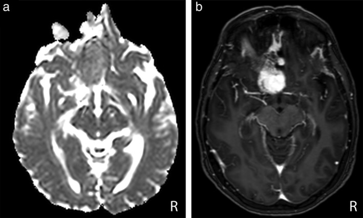
Field strength/sequence: 1.5 T, 3.0 T postcontrast enhanced T1 W (PCT1 W), apparent diffusion coefficient (ADC) maps (b values of 0, 500, and 1000 s/mm2 ).
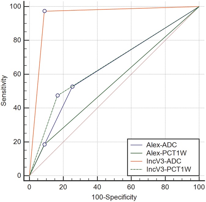
Assessment: WHO Grade II and WHO Grade III meningiomas were considered a single category. The diagnostic accuracy of the pretrained Inception-V3 and AlexNet DCNNs was tested on ADC maps and PCT1 W images separately. Receiver operating characteristic curves (ROC) and area under the curve (AUC) were used to asses DCNN performance.
Statistical test: Leave-one-out cross-validation.
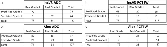
Results: The application of the Inception-V3 DCNN on ADC maps provided the best diagnostic accuracy results, with an AUC of 0.94 (95% confidence interval [CI], 0.88-0.98). Remarkably, only 1/38 WHO Grade II-III and 7/79 WHO Grade I lesions were misclassified by this model. The application of AlexNet on ADC maps had a low discriminating accuracy, with an AUC of 0.68 (95% CI, 0.59-0.76) and a high misclassification rate on both WHO Grade I and WHO Grade II-III cases. The discriminating accuracy of both DCNNs on postcontrast T1 W images was low, with Inception-V3 displaying an AUC of 0.68 (95% CI, 0.59-0.76) and AlexNet displaying an AUC of 0.55 (95% CI, 0.45-0.64).
Data conclusion: DCNNs can accurately discriminate between benign and atypical/anaplastic meningiomas from ADC maps but not from PCT1 W images.
Level of evidence: 2 Technical Efficacy: Stage 2 J. Magn. Reson. Imaging 2019;50:1152-1159.
Keywords: apparent diffusion coefficient; deep learning; grading; meningioma; postcontrast.
© 2019 The Authors. Journal of Magnetic Resonance Imaging published by Wiley Periodicals, Inc. on behalf of International Society for Magnetic Resonance in Medicine.
Figures
References
Publication types
MeSH terms
LinkOut - more resources
Full Text Sources
Medical
Research Materials
