Comparative Study
. 2019 Jul;24(4):e12647.
doi: 10.1111/anec.12647. Epub 2019 Mar 21.

Role of spatial dispersion of repolarization in reentry around a functional core versus reentry around a fixed anatomical core

Affiliations
Comparative Study

Role of spatial dispersion of repolarization in reentry around a functional core versus reentry around a fixed anatomical core

Herman D Himel et al. Ann Noninvasive Electrocardiol. 2019 Jul.

Abstract

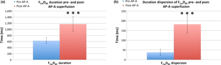
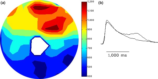
Introduction: Successful initiation of spiral wave reentry in the neonatal rat ventricular myocyte (NRVM) monolayer implicitly assumes the presence of spatial dispersion of repolarization (DR), which is difficult to quantify. We recently introduced a NRVM monolayer that utilizes anthopleurin-A to impart a prolonged plateau to the NRVM action potential. This was associated with a significant degree of spatial DR that lends itself to accurate quantification.

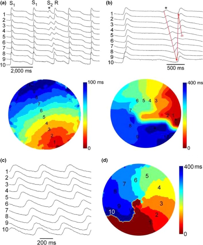
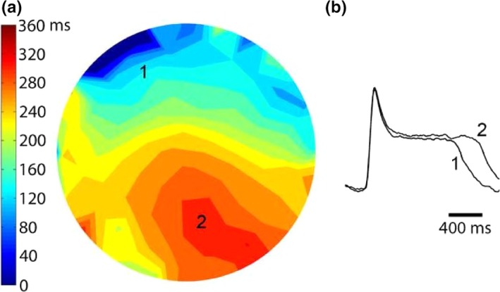
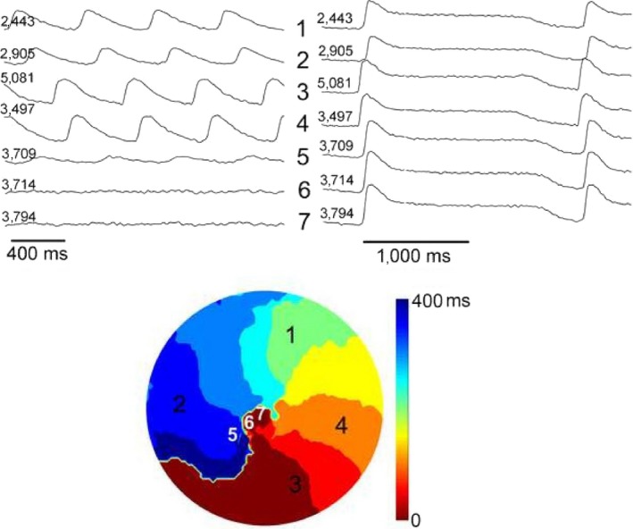
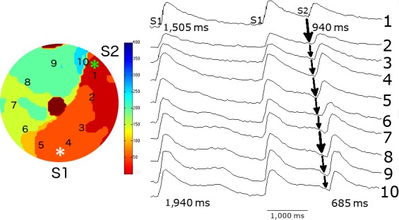
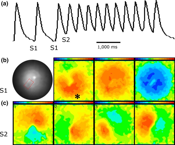
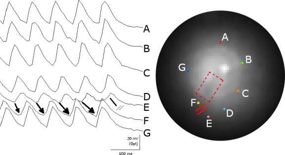
Methods and results: We utilized the monolayer and fluorescence optical mapping of intracellular calcium transients (FCai ) to systematically study and compare the contribution of spatial dispersion of the duration of FCai (as a surrogate of DR) to induction of spiral wave reentry around a functional core versus reentry around a fixed anatomical obstacle. We show that functional reentry could be initiated by a premature stimulus acting on a substrate of spatial DR resulting in a functional line of propagation block. Subsequent wave fronts circulated around a central core of functional obstacle created by sustained depolarization from the circulating wave front. Both initiation and termination of spiral wave reentry around an anatomical obstacle consistently required participation of a region of functional propagation block. This region was similarly based on spatial DR. Spontaneous termination of spiral wave reentry also resulted from block in the functional component of the circuit obstacle, usually preceded by beat-to-beat slowing of propagation.

Conclusions: The study demonstrates the critical contribution of DR to spiral wave reentry around a purely functional core as well as reentry around a fixed anatomical core.

Keywords: anthopleurin-A; dispersion of repolarization; neonatal rat ventricular myocyte monolayer; spiral wave reentry.


Conflict of interest statement

There is no conflict of interest.

Figures

Figure 1
Figure 1
Graphical representation of the FCaiD85 duration (left panel) and duration dispersion (right panel) pre‐ and post‐AP‐A superfusion with the monolayer stimulated at 0.5 Hz. Before superfusion, the FCaiD85 duration and duration dispersion in the monolayer were 626 ± 98 and 38 ± 16 ms (n = 6), respectively, and postperfusion both parameters significantly increased to 1,174 ± 255 and 182 ± 46 ms (n = 8)
Figure 2
Figure 2
Panels (a) and (b): Initiation of a single spiral wave reentry by an S1S2 protocol. The figure illustrates the last two beats of a series of 10 S1S1 stimulations at 0.5 Hz from a site close to the bottom of the optical field. The preparation was perfused with 4 nM AP‐A, resulting in prolongation of FCaiD85 to 1,500–1,600 ms. An S2 stimulus was applied to a site close to the right edge of the optical field at a CL of 1,250 ms (marked by asterisk) and induced a single reentrant spiral wave (R). Panel (a) shows 10 selected FCai potentials to illustrate the pathway of the FCai propagation wave of the S2 stimulus. Panel (b) illustrates an expanded view of the last S1 stimulus, the S2 stimulus, and the single spiral wave reentry. The isochronal map of both the S1 and S2 stimuli is shown underneath. The S1 map is drawn at 10 ms isochrones while the S2 map is drawn at 40 ms isochrones. The S1 stimulus resulted in a smooth propagation wave from the bottom to the top of the optical field at an average conduction velocity of 15 cm/s. On the other hand, the S2 stimulus failed to propagate to the bottom right half of the optical field resulting in a line of functional block that extended from the right edge to the middle of the optical field. The FCai wave front circulated around the left side edge of the line of block. However, the circulating wave front failed to propagate through the bottom side of the line of block, thus resulting in a single reentrant cycle. The expanded recordings in panel (b) illustrate both propagation of the circulating wave front (red arrow) and its final failure of propagation at the lower side of the line of functional block (double red bars). Panels (c) and (d) illustrate FCai signals and an isochronal map of sustained spiral wave reentry that was obtained 30 s following the recording in Panel (a) utilizing a similar S1S2 protocol. The spiral rotated at a CL of 400 ms around a 2 mm diameter core in the center of the optical field at approximately the same site as the left side end of the line of functional propagation block during the initiation of reentry in figure (a)
Figure 3
Figure 3
Panel (a) illustrates the repolarization map of the S1 beat that preceded the onset of spiral wave reentry shown in Figure 2. Isochrones of FCaiD85 were mapped at 40 ms duration. The line of functional propagation block around which the spiral wave reentry circulated developed at zones of crowded isochrones reflecting spatial dispersion of FCaiD85. Panel (b) shows two representative FCai signals recorded from two pixels in isochrones 160 and 360 ms and illustrate a 200 ms difference in FCaiD85
Figure 4
Figure 4
Illustrates the nature of FCai potentials in the core of the spiral wave reentry. The left panel illustrates selective FCai signals during sustained spiral wave reentry. The right panel illustrates the same signals during stimulation at 0.5 Hz. During reentry, there was a 2 mm diameter confluent zone of steady‐state small oscillations (sites 6 and 7). The approach to this zone showed low amplitude signals (site 5). The core of the spiral is made of a small area of tissue, which is depolarized and refractory, thus constituting a functional obstacle. This area is functionally normal during regular stimulation as shown in the right panel
Figure 5
Figure 5
Panel (a) was obtained from a monolayer with a fixed anatomical obstacle and illustrates the FCaiD85 map of the S1 stimulus that preceded the onset of spiral wave reentry. The figure shows a zone of spatial dispersion of FCaiD85 that extends from the central anatomical obstacle to the edge of the monolayer along the 2 o'clock line. Panel (b) illustrates two representative recordings of FCai from opposite sides of the zone of crowded isochrones. The FCaiD85 was 1,505 and 1,940 ms, respectively
Figure 6
Figure 6
Initiation of spiral wave reentry by an S1S2 stimulation protocol from the monolayer with a central anatomical obstacle. The middle panel illustrates the last two beats of a series of 10 S1S1 stimuli at 0.5 Hz and their position is shown in the right panel. An S2 stimulus (marked by asterisk) was applied to a site close to the zone of functional spatial dispersion of FCaiD85 and induced a single reentrant spiral wave. The selected FCai signals of S2 stimulus illustrate the pathway of the FCai propagation wave. The isochronal map of the S2 stimulus is shown on the left panel. The S2 stimulus failed to propagate in a counterclockwise direction because of a continuous line of block that includes the central anatomical obstacle and a functional zone of block. The wave front instead circulated in a clockwise direction around the central anatomical obstacle but failed to propagate through the functional line of block at the 1 o'clock zone, thus resulting in a single reentrant cycle. The figure illustrates the possible electrophysiological mechanism of the failure of the counterclockwise wave front to break through the line of functional block. The FCaiD85 of the two S1 signals on each side of the line of functional block were 1,505 and 1,940 ms. The duration of the closely coupled S2 signal at both sites showed significant shortening as expected. However, there was relatively more shortening of the FCaiD85 bottom signal. This resulted in a shorter duration of the FCaiD85 bottom S2 signal on the left side of the functional block compared to the FCaiD85 top S2 signal on the right side of the line of block. Repetitive reentry would have required a longer duration of the FCaiD85 S2 signal of the left side of the line of functional block compared to the signal on the right side of the block
Figure 7
Figure 7
The figure illustrates the initiation and spontaneous termination of spiral wave reentry from a different monolayer with a fixed anatomical core. Panel (a) shows FCai recordings from one pixel that illustrates the last three of a series of 10 S1 stimuli at a CL of 1 Hz followed by an S2 stimulus that initiated a train of nine cycles of spiral waves that terminated spontaneously. Panel (b) consists of four sections. The section on the left is a low power fluoroscopic image of the monolayer that illustrates the anatomical obstacle that was created in the middle of the monolayer by scraping the cells from a 2 × 3 mm section (visible as a darker zone in the fluoroscopic image compared to the rest of the monolayer and is delineated by dotted line). The remaining three sections illustrate the FCai wave front of the S1 stimulus. The propagation of FCai signal is represented in red while the absence of FCai signals in the monolayer is represented in blue. The site of the S1 stimulus is marked by an asterisk. The advancing FCai wave front circulated around the anatomical obstacle (section 2) and propagated quickly within less than 40 ms to the rest of the monolayer (section 3) before the monolayer became quiet during the interval between successive S1 stimuli (labeled blue). Panel (c) illustrates sequential recordings of the clockwise rotating FCai wave front (consecutive sections 1 to 4) representing spiral wave reentry
Figure 8
Figure 8
The mechanism of spontaneous termination of the spiral wave reentry. The figure illustrates seven FCai signals from selected pixels around the spiral wave front (marked by different colors on the fluoroscopic image of the monolayer and labeled A to G). The recording illustrates the sequential rotation of the FCai wave front around the anatomical obstacle. The termination of the spiral wave reentry occurred at a site located between the E and F traces (marked by a line with double cross bars). The gradual lengthening of the arrows drawn between the E and F traces represents gradual slowing of propagation of the FCai wave front between these two sites before propagation failure terminated the reentrant activation. The conduction delay and terminal block of reentrant activation took place in the normal section of the monolayer outside the fixed anatomical obstacle. Thus, termination of the circulating wave front has to occur at a continuous line of block that consists of the anatomical fixed obstacle and a functional line of block that extends the electrophysiological barrier to the edge of the monolayer

Similar articles

Cited by

References

    1. Agladze, K. , Kay, M. W. , Krinsky, V. , & Sarvazyan, N. (2007). Interaction between spiral and paced waves in cardiac tissue. American Journal of Physiology‐Heart and Circulatory Physiology, 293(1), H503–H513. 10.1152/ajpheart.01060.2006 - DOI - PMC - PubMed
    1. Antezelevich, C. (2001). Heterogeneity of cellular repolarization in LQTS: The role of M cells. European Heart Journal Supplements, 3(Suppl. K), K2–K16. 10.1016/S1520-765X(01)90001-X - DOI
    1. Boutjdir, M. , Restivo, M. , Wei, Y. , Stergiopoulos, K. , & El‐Sherif, N. (1994). Early afterdepolarizations formation in cardiac myocytes: Analysis of phase plane patterns, action potential, and membrane currents. Journal of Cardiovascular Electrophysiology, 5(7), 609–620. 10.1111/j.1540-8167.1994.tb01302.x - DOI - PubMed
    1. El‐Sherif, N. , Caref, E. B. , Yin, H. , & Restivo, M. (1996). The electrophysiological mechanism of ventricular tachyarrhythmias in the long QT syndrome. Tridimensional mapping of activation and recovery patterns. Circulation Research, 79(3), 474–492. 10.1161/01.res.79.3.474 - DOI - PubMed
    1. El‐Sherif, N. , Fozzard, H. A. , & Hanck, D. (1992). Dose‐dependent modulation of the cardiac sodium channel by the sea anemone toxin ATX‐II. Circulation Research, 70(2), 285–301. 10.1161/01.RES.70.2.285 - DOI - PubMed

Publication types

Substances