. 2019 May;52(3):e12601.
doi: 10.1111/cpr.12601. Epub 2019 Mar 21.

P34HB electrospun fibres promote bone regeneration in vivo

Affiliations

P34HB electrospun fibres promote bone regeneration in vivo

Na Fu et al. Cell Prolif. 2019 May.

Abstract

Objective: Bone tissue engineering was introduced in 1995 and provides a new way to reconstruct bone and repair bone defects. However, the design and fabrication of suitable bionic bone scaffolds are still challenging, and the ideal scaffolds in bone tissue engineering should have a three-dimensional porous network, good biocompatibility, excellent biodegradability and so on. The purpose of our research was to investigate whether a bioplasticpoly3-hydroxybutyrate4-hydroxybutyrate (P34HB) electrospun fibre scaffold is conducive to the repair of bone defects, and whether it is a potential scaffold for bone tissue engineering.

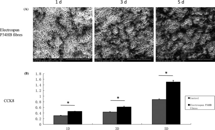
Materials and methods: The P34HB electrospun fibre scaffolds were prepared by electrospinning technology, and the surface morphology, hydrophilicity, mechanical properties and cytological behaviour of the scaffolds were tested. Furthermore, a calvarial defect model was created in rats, and through layer-by-layer paper-stacking technology, the P34HB electrospun fibre scaffolds were implanted into the calvarial defect area and their effect on bone repair was evaluated.

Results: The results showed that the P34HB electrospun fibre scaffolds are interwoven with several fibres and have good porosity, physical properties and chemical properties and can promote cell adhesion and proliferation with no cytotoxicity in vitro. In addition, the P34HB electrospun fibre scaffolds can promote the repair of calvarial defects in vivo.

Conclusions: These results demonstrated that the P34HB electrospun fibre scaffold has a three-dimensional porous network with good biocompatibility, excellent biosafety and ability for bone regeneration and repair; thus, the P34HB electrospun fibre scaffold is a potential scaffold for bone tissue engineering.

Keywords: P34HB; bone marrow mesenchymal stem cells; bone tissue engineering; calvarial defects; electrospinning.


Figures

Figure 1
Figure 1
Characterization of P34HB electrospun fibres scaffold. A, Morphology of the scaffold evaluated by SEM (n = 3). The structure of the scaffold is interwoven by a number of fibres, which are arranged randomly and have many holes. B, Measurement of the water contact angle wetting behaviour of a water droplet on the scaffold (n = 3), the contact angle was determined at 116.5 ± 2.3°. C, Mechanical parameters determined from slices of the scaffold (10.0 × 5.0 × 0.7 m3) (n = 3). The maximum strength the scaffold could bear was 5 N, and the scaffold can be stretched to ~6 mm before breaking. D, The mechanical parameters of fibres were measured. The Young's modulus of randomly selected fibres was approximately 58.929 MPa, and the elongation at the breaking point was 26.887%
Figure 2
Figure 2
Compared with the control group, P34HB electrospun fibres scaffold can promote cell adhesion and proliferation. As shown by microscope observation, bone marrow mesenchymal stem cells (BMSCs) attached to the scaffold after seeding. As time goes on, the cell spreading area was the broader and the number of BMSCs was significantly increased in comparison to the control group
Figure 3
Figure 3
In vitro cell behaviour of the P34HB electrospun fibres scaffold. A, Cell morphologies of bone marrow mesenchymal stem cells (BMSCs) on the fibres, as shown by SEM (n = 3). BMSCs were seeded, attached, spread and proliferated within 5 days. B, Cell proliferation of BMSCs on the fibres and Petri dishes (n = 3). The results show that the proliferation rates within 5 days were significantly higher on the fibres than that found on the Petri dishes
Figure 4
Figure 4
Radiographical analysis of bone formation. Compared with the control group, the effect of the scaffold group on defect repair was better at both time points. There were more green areas in the control group and more red areas in the scaffold group, where green represents low mineralized fibrous tissue and red represents mineralized bone. At 8 weeks, the best results were achieved in the scaffold group, and the defect area was basically covered by red. Moreover, new bone‐like tissue grew from the periphery of the defect area into the centre
Figure 5
Figure 5
HE staining of cross‐sections of repaired calvarial defects at 4 and 8 weeks. The results of each group at 8 weeks were better than those at 4 weeks, and the P34HB group results were better than the results of the control group. In the control group, we observed only a large amount of connective tissue and a small amount of bone‐like tissue, 8 weeks after implantation, the P34HB scaffolds had a large number of new bone islands, some regenerated bone and bone‐like tissue had replaced the gradually degraded scaffold material, and the bony bridge was almost fully linked with clear, mature bone structures (bl, bone‐like tissue; ct, connective tissue; rb, regenerated bone; green arrows, layered scaffold structures)
Figure 6
Figure 6
The results of Masson's trichome staining were consistent with those of HE staining. The P34HB group results were better than the results of the control group at the two time points. In the control group, we observed only a large amount of connective tissue and a small amount of bone‐like tissue. The best results were obtained after 8 weeks of implantation when a large number of new bone islands could be seen, some regenerated bone and bone‐like tissue had replaced the gradually degraded scaffold material, and the bony bridge was almost fully linked with clear, mature bone structures (bl, bone‐like tissue; ct, connective tissue; rb, regenerated bone; green arrows: layered scaffold structures; black arrow, blood vessels)

Similar articles

Cited by

References

    1. Petite H, Viateau V, Bensaïd W, et al. Tissue‐engineered bone regeneration. Nat Biotechnol. 2000;18:959‐963. - PubMed
    1. Wang H, Li Y, Zuo Y, Li J, Ma S, Cheng L. Biocompatibility and osteogenesis of biomimetic nano‐hydroxyapatite/polyamide composite scaffolds for bone tissue engineering. Biomaterials. 2007;28:3338‐3348. - PubMed
    1. Stevens MM. Biomaterials for bone tissue engineering. Mater Today. 2008;11:18‐25.
    1. Bran GM, Stern‐Straeter J, Hörmann K, Riedel F, Goessler UR. Apoptosis in bone for tissue engineering. Arch Med Res. 2008;39:467‐482. - PubMed
    1. Cancedda R, Giannoni P, Mastrogiacomo M. A tissue engineering approach to bone repair in large animal models and in clinical practice. Biomaterials. 2007;28:4240‐4250. - PubMed