

Phenotypic distinctions between the nodose and jugular TRPV1-positive vagal sensory neurons in the cynomolgus monkey
- PMID: 30896676
- PMCID: PMC6481665
- DOI: 10.1097/WNR.0000000000001231
Phenotypic distinctions between the nodose and jugular TRPV1-positive vagal sensory neurons in the cynomolgus monkey
Abstract
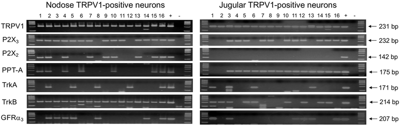
Vagal capsaicin-sensitive afferent C-fibers play an important role in the maintenance of visceral homeostasis and contribute to symptoms in visceral diseases. Based on their developmental origin two functionally distinct types of vagal C-fibers are recognized: those with neurons in the vagal nodose ganglia (derived from epibranchial placodes) and in the vagal jugular ganglia (from neural crest). Studies in nonprimate species demonstrated that the vagal nodose and jugular C-fibers differ in activation profile, neurotrophic regulation, and expression of neurotransmitters. We hypothesized that the expression of selected markers related to key phenotypic properties of vagal C-fibers in the cynomolgus monkey is similar to that reported in nonprimate species. We performed single-cell RT-PCR on nodose and jugular putative C-fiber (TRPV1-positive) neurons isolated from the cynomolgus monkey. We found that the expression of purinergic P2X receptors that underlie selective responsiveness of nodose C-fiber terminals to ATP was conserved in that P2X2 and P2X3 subunits were expressed in nodose, but only P2X3 subunit was expressed in jugular TRPV1-positive neurons. Also conserved was the preferential expression of neurotrophic receptor TrkB in the nodose and preprotachykinin-A in the jugular TRPV1-positive neurons. Several key distinctions in gene expression between nodose and jugular TRPV1-positive (C-fiber) neurons that have been noted in mice, rats, and guinea pigs, are conserved in the cynomolgus monkey. Our results support the translatability of distinct vagal C-fiber phenotypes to primates.
Conflict of interest statement
Conflicts of Interest and Source of Funding
Conflict of interest: none declared.
Figures
References
-
- Baker CV, Bronner-Fraser M. Vertebrate cranial placodes I. Embryonic induction. Dev Biol 2001; 232: 1–61. - PubMed
-
- Rhoton AL Jr., O’Leary JL, Ferguson JP. The trigeminal, facial, vagal, and glossopharyngeal nerves in the monkey. Afferent connections. Archives of neurology 1966; 14: 530–540. - PubMed
-
- Satoda T, Takahashi O, Uchida T, Mizuno N. An anterograde-retrograde labeling study of the carotid sinus nerve of the Japanese monkey (Macaca fuscata). Neuroscience research 1995; 22: 381–387. - PubMed
-
- Kummer W, Fischer A, Kurkowski R, Heym C. The sensory and sympathetic innervation of guinea-pig lung and trachea as studied by retrograde neuronal tracing and double-labelling immunohistochemistry. Neuroscience 1992; 49: 715–737. - PubMed
Publication types
MeSH terms
Substances
Grants and funding
LinkOut - more resources
Full Text Sources
Other Literature Sources
