. 2019 May;54(5):1567-1578.
doi: 10.3892/ijo.2019.4756. Epub 2019 Mar 19.

Oral mucosal mesenchymal stem cell‑derived exosomes: A potential therapeutic target in oral premalignant lesions

Affiliations

Oral mucosal mesenchymal stem cell‑derived exosomes: A potential therapeutic target in oral premalignant lesions

Wenwen Li et al. Int J Oncol. 2019 May.

Abstract

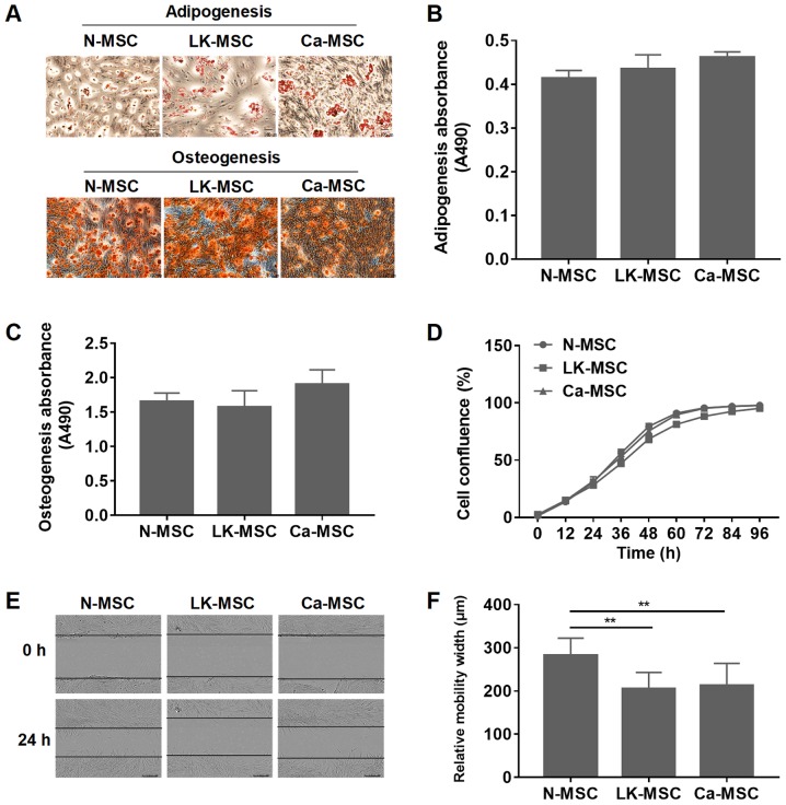
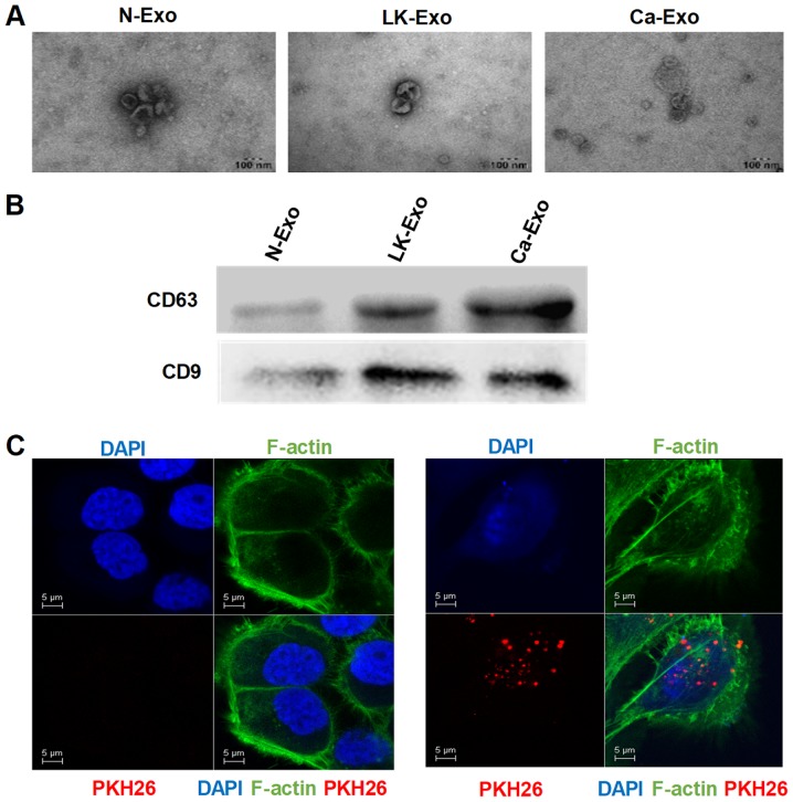
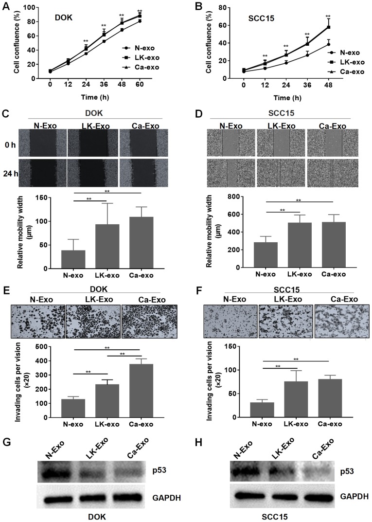
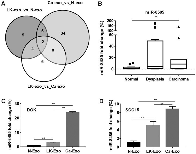
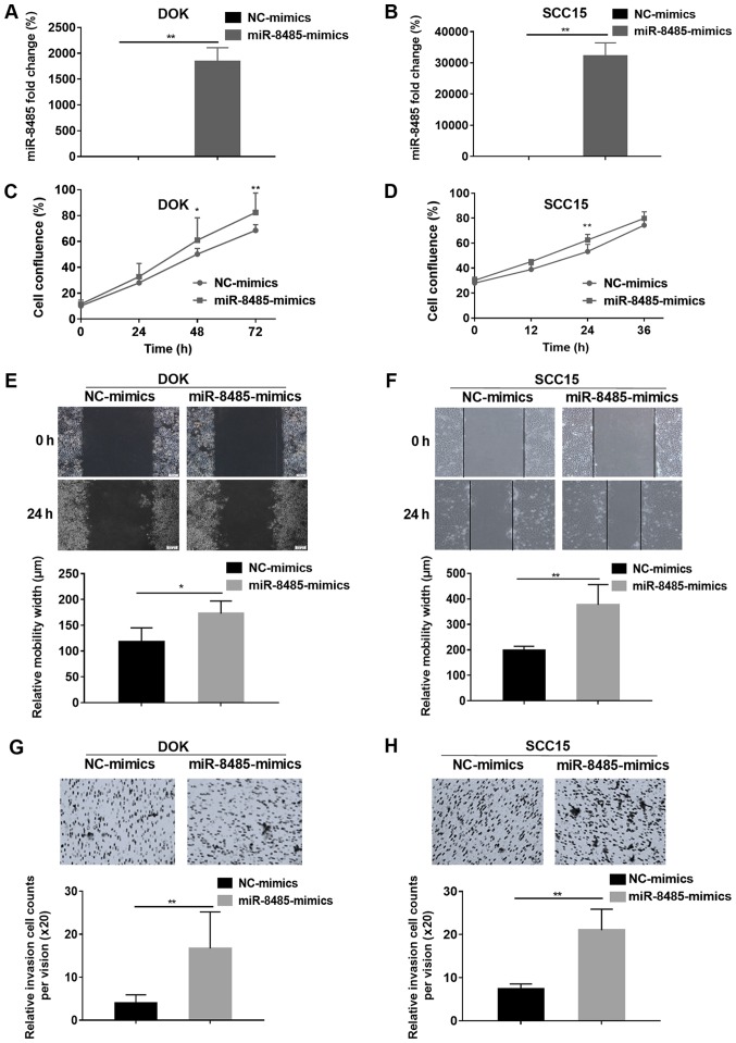
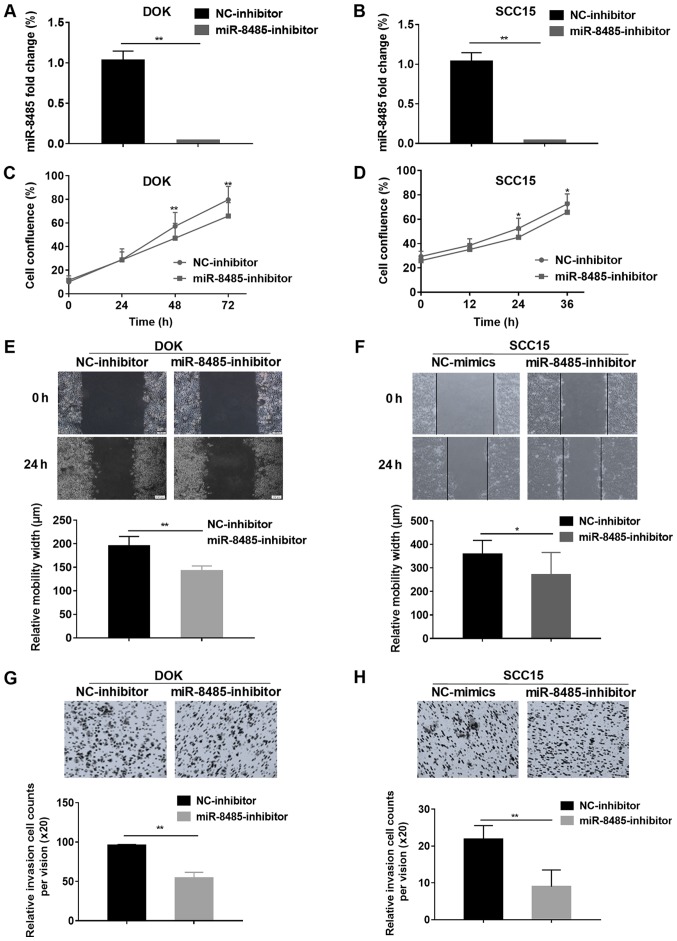
Emerging evidence indicates that mesenchymal stem cells (MSCs) serve an indispensable role in the tumor microenvironment. However, whether MSCs participate in the development of oral carcinogenesis remains unclear. The present study isolated MSCs from clinical tissues and investigated the differences of MSCs derived from normal oral mucosa (N‑MSC), oral leukoplakia with dysplasia (LK‑MSC) and oral carcinoma (Ca‑MSC). The results revealed that the LK‑MSCs exhibited reduced proliferation and migration, compared with the N‑MSCs and Ca‑MSCs. Furthermore, it was demonstrated that the exosomes secreted by LK‑MSCs have significant roles in promoting proliferation, migration and invasion in vitro, which was similar to the Ca‑MSC‑derived exosomes. The promoting effect was also demonstrated in a 3D coculture model. When the secretion of exosomes was blocked, the promoting effect of LK‑MSCs was reversed. Based on a microarray analysis of MSC‑derived exosomes, microRNA‑8485 (miR‑8485) was identified to be ectopically expressed. The exosomal miR‑8485 was capable of promoting the proliferation, migration and invasion of tumor cells. Therefore, the present study highlights the significance of MSC‑derived exosomes and exosomal miR‑8485 in premalignant lesions and carcinogenesis. Intervention with the secretion of MSC‑derived‑exosomes may be an innovative strategy to retard the carcinogenesis.


Figures

Figure 1
Figure 1
Characteristics of the N-MSCs, LK-MSCs and Ca-MSCs. (A) Adipogenic and osteogenic differentiation were assessed. Representative images with a magnification of ×10 are depicted. (B) Quantitative analysis of adipogenic differentiation was calculated using the absorbance value at 490 nm. (C) Quantitative analysis of osteogenic differentiation was calculated using the absorbance value at 490 nm. (D) MSCs were cultured and the cell confluence was analyzed with an InCucyte monitoring microscope. (E and F) The relative migration width was determined by a wound healing assay. Representative images with a magnification of ×10 are depicted. **P<0.01. MSC, mesenchymal stem cell; N-MSC, MSCs derived from normal oral mucosa; LK-MSC, MSCs derived from oral leukoplakia with dysplasia; Ca-MSC, MSCs derived from oral carcinoma.
Figure 2
Figure 2
Identification of exosomes. (A) Exosomes were isolated from MSCs and observed using transmission electron microscopy. (B) The western blotting revealed that the CD63 and CD9 proteins were expressed in the exosomes. (C) Confocal microscopy was used to identify the uptake of PKH26-labeled exosomes, which were secreted by N-MSC and internalized in the cytoplasm. N-exo, exosomes secreted by MSCs derived from normal oral mucosa; LK-exo, exosomes secreted by MSCs derived from oral leukoplakia with dysplasia; Ca-exo, exosomes secreted by MSCs derived from oral carcinoma; MSCs, mesenchymal stem cells; CD63, cluster of differentiation 63.
Figure 3
Figure 3
Effects of MSC-derived exosomes on epithelial cells. (A) The effect of the exosomes on proliferation was determined by the confluence of DOK cells. (B) The effect of the exosomes on proliferation was determined by the confluence of SCC15 cells. (C) The relative migration width of DOK cells was determined by a wound healing assay. Representative images with a magnification of ×10 are depicted. (D) The relative migration width of SCC15 cells was determined by a wound healing assay. Representative images with a magnification of ×10 are depicted. (E) The invasion ability of DOK cells treated with exosomes was assessed by a Matrigel cell invasion assay. Representative images with a magnification of ×20 are depicted. (F) The invasion ability of SCC15 cells treated with exosomes was assessed by a Matrigel cell invasion assay. Representative images with a magnification of ×20 are depicted. (G) The expression of p53 was assessed by western blotting for DOK cells. (H) The expression of p53 was assessed by western blotting for SCC15 cells. **P<0.01, compared with N-exo. N-exo, exosomes secreted by MSCs derived from normal oral mucosa; LK-exo, exosomes secreted by MSCs derived from oral leukoplakia with dysplasia; Ca-exo, exosomes secreted by MSCs derived from oral carcinoma; MSCs, mesenchymal stem cells.
Figure 4
Figure 4
SCC15 cells cocultured with MSCs in a 3D coculture model. MSCs and SCC15 cells were cocultured in a 3D coculture model for 5 days. The cocultures were assessed with H&E and immunohistochemical staining. Representative images with a magnification of ×20 are depicted. MSCs, mesenchymal stem cells. MSC, mesenchymal stem cell; N-MSC, MSCs derived from normal oral mucosa; LK-MSC, MSCs derived from oral premalignant lesion; Ca-MSC, MSCs derived from oral carcinoma; H&E, haematoxylin and eosin.
Figure 5
Figure 5
Expression of exosomal miR-8485 was gradually elevated. (A) The miRNA from the N-exo, LK-exo and Ca-exo groups was analyzed. Among the miRNAs measured with fold change >2, the expression levels of miR-4433a and miR-8485 were both changed. (B) The expression of miR-8485 in the clinical tissues was assessed by reverse transcription-quantitative polymerase chain reaction. The box represents the interquartile range (25-75th percentile) and the line within this box is the median value. The bottom and top bars of the whisker plot indicate the 10th and 90th percentiles, respectively. (C) DOK cells treated with LK-exo and Ca-exo exhibited elevated expression of miR-8485. (D) SCC15 cells treated with LK-exo and Ca-exo exhibited elevated expression of miR-8485. *P<0.05 and **P<0.01. miR, microRNA; N-exo, exosomes from mesenchymal stem cells derived from normal oral mucosa; LK-exo, exosomes from mesenchymal stem cells derived from oral premalignant lesion; Ca-exo, exosomes from mesenchymal stem cells derived from oral carcinoma.
Figure 6
Figure 6
Overexpression of miR-8485 promotes the migration and invasion of DOK and SCC15 cells. (A) The miR-8485 mimics and mimics control (NC-mimics) were transfected into DOK cells, and the efficiency was assessed by reverse transcription-quantitative polymerase chain reaction. (B) The miR-8485 mimics and mimics control (NC-mimics) were transfected into SCC15 cells, and the efficiency was assessed by reverse transcription-quantitative polymerase chain reaction. (C) The effect of the miR-8485 mimics on proliferation was analyzed by assessing cell confluence of DOK cells. (D) The effect of the miR-8485 mimics on proliferation was analyzed by assessing cell confluence of SCC15 cells. (E) The relative migration width of the DOK cells following overexpression of miR-8485 was determined with a wound healing assay. Representative images with a magnification of ×10 are depicted. (F) The relative migration width of the SCC15 cells following overexpression of miR-8485 was determined with a wound healing assay. Representative images with a magnification of ×10 are depicted. (G) The Matrigel invasion assay indicated the invasion ability of the DOK cells. Representative images with a magnification of ×20 are depicted. (H) The Matrigel invasion assay indicated the invasion ability of the SCC15 cells. Representative images with a magnification of ×20 are depicted. *P<0.05 and **P<0.01, compared with NC groups. NC, negative control; miR, microRNA.
Figure 7
Figure 7
miR-8485 inhibitor suppresses the migration and invasion of DOK and SCC15 cells. (A) miR-8485 expression was detected by reverse transcription-quantitative polymerase chain reaction after miR-8485 inhibitor was transfected into the DOK cells. (B) miR-8485 expression was detected by reverse transcription-quantitative polymerase chain reaction after miR-8485 inhibitor was transfected into the SCC15 cells. (C) The effect of the miR-8485 inhibitor on proliferation was analyzed by the cell confluence of DOK cells. (D) The effect of the miR-8485 inhibitor on proliferation was analyzed by the cell confluence of SCC15 cells. (E) The wound healing assay revealed that compared with the NC-inhibitor, the miR-8485 inhibitor suppressed the migration of the DOK cells. Representative images with a magnification of ×10 are depicted. (F) The wound healing assay revealed that compared with the NC-inhibitor, the miR-8485 inhibitor suppressed the migration of the SCC15 cells. Representative images with a magnification of ×10 are depicted. (G) The Matrigel invasion assay indicated that compared with the NC-inhibitor, the miR-8485 inhibitor suppressed the invasion ability of the DOK cells. Representative images with a magnification of ×20 are depicted. (H) The Matrigel invasion assay indicated that compared with the NC-inhibitor, the miR-8485 inhibitor suppressed the invasion ability of the SCC15 cells. Representative images with a magnification of ×20 are depicted. *P<0.05 and **P<0.01, compared with NC groups. NC, negative control; miR, microRNA.

Similar articles

Cited by

References

    1. Bray F, Ferlay J, Soerjomataram I, Siegel RL, Torre LA, Jemal A. Global cancer statistics 2018: GLOBOCAN estimates of incidence and mortality worldwide for 36 cancers in 185 countries. CA Cancer J Clin. 2018;68:394–424. doi: 10.3322/caac.21492. - DOI - PubMed
    1. Bouquot JE, Whitaker SB. Oral leukoplakia - rationale for diagnosis and prognosis of its clinical subtypes or 'phases'. Quintessence Int. 1994;25:133–140. - PubMed
    1. Kao SY, Mao L, Jian XC, Rajan G, Yu GY. Expert consensus on the detection and screening of oral cancer and precancer. Chin J Dent Res. 2015;18:79–83. - PubMed
    1. van der Waal I, Schepman KP, van der Meij EH, Smeele LE. Oral leukoplakia: A clinicopathological review. Oral Oncol. 1997;33:291–301. doi: 10.1016/S1368-8375(97)00002-X. - DOI - PubMed
    1. Vohra F, Al-Kheraif AA, Qadri T, Hassan MI, Ahmed A, Warnakulasuriya S, Javed F. Efficacy of photodynamic therapy in the management of oral premalignant lesions. A systematic review. Photodiagn Photodyn Ther. 2015;12:150–159. doi: 10.1016/j.pdpdt.2014.10.001. - DOI - PubMed

MeSH terms