. 2019 May;19(5):3743-3755.
doi: 10.3892/mmr.2019.10037. Epub 2019 Mar 14.

β‑estradiol alleviates hypertension‑ and concanavalin A‑mediated inflammatory responses via modulation of connexins in peripheral blood lymphocytes

Affiliations

β‑estradiol alleviates hypertension‑ and concanavalin A‑mediated inflammatory responses via modulation of connexins in peripheral blood lymphocytes

Xin Ni et al. Mol Med Rep. 2019 May.

Abstract

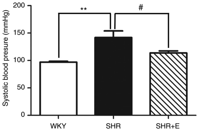
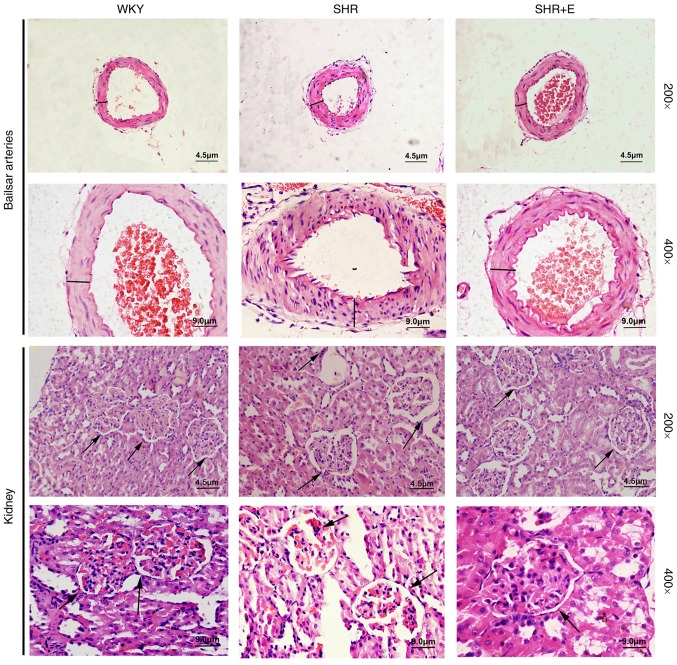
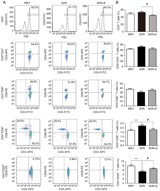
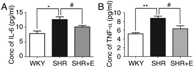
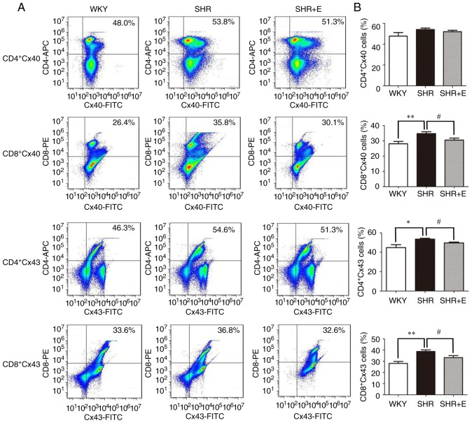
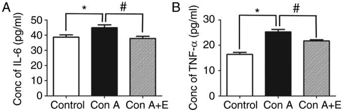
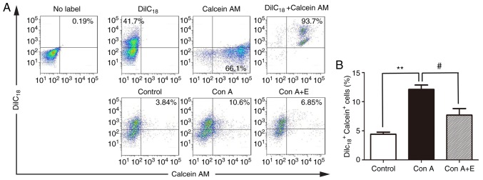
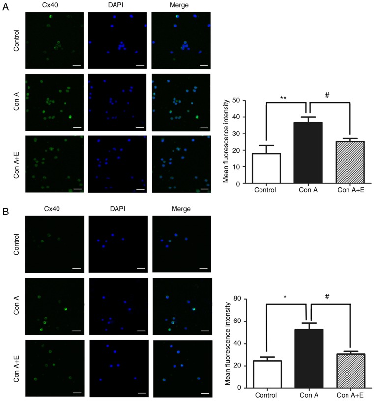
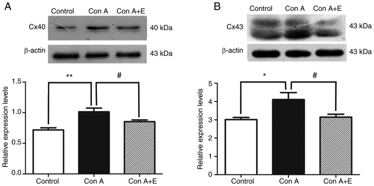
Gap junctions (GJs) formed by connexins (Cxs) in T lymphocytes have been reported to have important roles in the T lymphocyte‑driven inflammatory response and hypertension‑mediated inflammation. Estrogen has a protective effect on cardiovascular diseases, including hypertension and it attenuates excessive inflammatory responses in certain autoimmune diseases. However, the mechanisms involved in regulating the pro‑inflammatory response are complex and poorly understood. The current study investigated whether β‑estradiol suppresses hypertension and pro‑inflammatory stimuli‑mediated inflammatory responses by regulating Cxs and Cx‑mediated GJs in peripheral blood lymphocytes. Male, 16‑week‑old spontaneously hypertensive rats (SHR) and Wistar‑Kyoto rats (WKY) rats were randomly divided into the following three groups: WKY rats, vehicle (saline)‑treated SHRs, and β‑estradiol (20 µg/kg/day)‑treated SHRs. β‑estradiol was administered subcutaneously for 5 weeks. Hematoxylin and eosin staining was performed to evaluate target organ injury. Flow cytometry and ELISA were used to measure the populations of T lymphocyte subtypes in the peripheral blood, and expression of Cx40/Cx43 in T cell subtypes, and pro‑inflammation cytokines levels, respectively. ELISA, a dye transfer technique, immunofluorescence and immunoblotting were used to analyze the effect of β‑estradiol on pro‑inflammatory cytokine secretion, Cx‑mediated GJs and the expression of Cxs in concanavalin A (Con A)‑stimulated peripheral blood lymphocytes isolated from WKY rat. β‑estradiol significantly decreased blood pressure and inhibited hypertension‑induced target organ injury in SHRs. Additionally, β‑estradiol treatment significantly improved the immune homeostasis of SHRs, as demonstrated by the decreased percentage of cluster of differentiation (CD)4+/CD8+ T‑cell subset ratio, reduced serum levels of pro‑inflammatory cytokines and increased the percentage of CD4+CD25+ T cells. β‑estradiol also markedly reduced the expression of Cx40/Cx43 in T lymphocytes from SHRs. In vitro, β‑estradiol significantly suppressed the production of pro‑inflammatory cytokines, reduced communication via Cx‑mediated gap junctions and decreased the expression of Cx40/Cx43 in Con A‑stimulated lymphocytes. These results indicate that β‑estradiol attenuates inflammation and end organ damage in hypertension, which may be partially mediated via downregulated expression of Cxs and reduced function of Cx‑mediated GJ.


Figures

Figure 1.
Figure 1.
Effects of β-estradiol supplement on tail systolic arterial pressure in SHR. Tail systolic arterial pressure of the SHR group significantly increased in comparison with the WKY group. Compared with SHR without β-estradiol treatment, chronic subcutaneous injection of 20 µg/kg/day β-estradiol for 5 weeks significantly attenuated hypertension in the SHR + E group. Values are presented as the mean ± standard error, n=6. **P<0.01 (P=0.0061), WKY vs. SHR and #P<0.05 (P=0.0447), SHR vs SHR + E. SHR, spontaneously hypertensive rat; WKY, Wistar-Kyoto rats; SHR + E, SHR + β-estradiol.
Figure 2.
Figure 2.
β-estradiol supplement attenuates vascular remodeling of BA, target organ damage and inflammatory cell infiltration in BA and kidney tissues of SHR. Representative pictures of hematoxylin and eosin staining of renal and arterial sections indicated the protective role of β-estradiol on target organ damage. The two highlighted black lines represent the label of basilar arteries and kidney. The arrows indicate atrophy of the glomerulus, infiltration of inflammatory cell into part of renal interstitium. Magnification, ×200 (scale bar, 4.5 µm) or ×400 (scale bar, 9.0 µm). n=6. BA, basilar arteries; SHR, spontaneously hypertensive rat; WKY, Wistar-Kyoto rats; SHR + E, SHR + β-estradiol.
Figure 3.
Figure 3.
β-estradiol supplement reverses the changes in the percentages of various T lymphocyte subpopulations in peripheral circulation of SHR. The percentage of CD3+, CD3+CD4+, CD3+CD8+, CD4+ CD25+ T cells was analyzed by flow cytometry. (A) Flow cytometry dot plots represent the percentages of circulating T lymphocytes subtypes in the peripheral blood of SHR and WKY rats. Decrease in the CD3+ T cell population and CD4+/CD8+ T cell subset ratios, and increased percentage of CD4+CD25+ T cells were observed following β-estradiol treatment in SHR. (B) Bar graphs represent the percentage of various T cell subpopulations and CD4+/CD8+ T cell subset ratios. All numerical data are displayed as the mean ± standard error (n=6/group). **P<0.01 vs. WKY group and SHR group without any drug treatment. #P<0.05 SHR group without any drug treatment vs. SHR group given β-estradiol. SHR, spontaneously hypertensive rat; WKY, Wistar-Kyoto rats; CD, cluster of differentiation; SHR + E, SHR + β-estradiol; FITC, fluorescein isothiocyanate; PE, phycoerythrin; APC, allophycocyanin; FSC, forward scatter.
Figure 4.
Figure 4.
β-estradiol supplement attenuates hypertension induced cytokine production in vivo. Concentrations of (A) IL-6 and (B) TNF-α in serum of WKY, SHR and SHR received β-estradiol treatment. Data are presented as the mean ± standard error of three independent experiments (n=6/group). *P<0.05 and **P<0.01 vs. the WKY group and SHR group without any drug treatment. #P<0.05, SHR group without any drug treatment vs. SHR group given β-estradiol. TNF-α, tumor necrosis factor-α; IL, interleukin; WKY, Wistar-Kyoto rats; SHR, spontaneously hypertensive rat; SHR + E, SHR + β-estradiol.
Figure 5.
Figure 5.
β-estradiol supplement reduces the percentages of Cx40- and Cx43-expressing CD4+ and CD8+ T cells in SHR. (A) Representative dot plot and (B) collated data presenting percentages of Cx40- and Cx43-positive CD4+ and CD8+ T cells in each group. Data are presented as the mean ± standard error of three independent experiments (n=6/group). *P<0.05 and **P<0.01, WKY group vs. SHR group without any drug treatment; #P<0.05, SHR group without any drug treatment vs. SHR group with β-estradiol treatment. Cx, connexin; SHR, spontaneously hypertensive rat; WKY, Wistar-Kyoto rats; SHR + E, SHR + β-estradiol; CD, cluster of differentiation; FITC, fluorescein isothiocyanate; PE, phycoerythrin; APC, allophycocyanin.
Figure 6.
Figure 6.
β-estradiol pre-treatment decreased Con A-induced secretion of pro-inflammatory cytokines in the culture supernatant of peripheral blood lymphocytes stimulated with Con A. Contents of (A) IL-6 and (B) TNF-α in the culture supernatant of unstimulated, Con A stimulated and β-estradiol treated peripheral blood lymphocytes. Data are presented as the mean ± standard error (n=5). *P<0.05, control group vs. the Con A-stimulated group; #P<0.05, Con A-stimulated group vs. the β-estradiol treated group. Each figure is representative of three independent experiments. Con A, concanavalin A; Con A + E, concanavalin A + β-estradiol; IL, interleukin; TNF, tumor necrosis factor.
Figure 7.
Figure 7.
β-estradiol supplement suppresses the function of gap junctions in Con A stimulated peripheral blood lymphocytes. (A) Control experiments of DiIC18 or calcein AM single-stained cells and DiIC18-calcein double-stained cells were performed in parallel; DiIC18+-calcein+-positive fluorescent cells expressed as a represent percentage of the total number of peripheral blood lymphocytes. (B) Mean percentages of DiIC18+-calcein+-double-positive cells of three independent experiments ± standard error from five samples/group. **P<0.01, control group vs. Con A-stimulated group; #P<0.05, Con A-stimulated group vs. E-treated group. Con A, concanavalin A; calcein AM, calcein acetoxymethyl ester; E, β-estradiol.
Figure 8.
Figure 8.
β-estradiol pre-treatment decreased Cx40/Cx43 expression in peripheral blood lymphocytes stimulated with Con A. Representative immunofluorescence images of (A) Cx40 and (B) Cx43 (magnification, ×630; scale bar, 16 µm) to determine their localization and expression in the plasma membrane and cytosol. These images presenting the changes of Cx40 and Cx43 expression when Con A stimulated peripheral blood lymphocytes were pretreated with β-estradiol. Bar graphs presenting densitometric analysis of mean immunofluorescent intensity of Cx40 and Cx43 in various groups. Each figure is representative of three independent experiments. Each column represents the mean ± standard error from five samples/group. *P<0.05 or **P<0.01, control group vs. Con A-stimulated group; #P<0.05, Con A-stimulated group vs. E-treated group. Cx, connexin; Con A, concanavalin A; DAPI, 4′,6-diamidino-2-phenylindole; E, β-estradiol.
Figure 9.
Figure 9.
β-estradiol pre-treatment inhibits Con A-induced protein expression of Cx40 and Cx43 in peripheral blood lymphocytes. The immunoblot analysis of (A) Cx40 and (B) Cx43 in peripheral blood lymphocytes treated with Con A and Con A + β-estradiol. The bar graph presents the densitometric analysis of relative expression of (A) Cx40 and (B) Cx43 normalized to β-actin level. Results are presented as the mean ± standard error of five samples/group. *P<0.05 and **P<0.01, control group vs. Con A-stimulated group; #P<0.05, Con A-stimulated group vs. β-estradiol treated group. Each figure is representative of three independent experiments. Con A, concanavalin A; Cx, connexin.

Similar articles

Cited by

References

    1. Abais-Battad JM, Dasinger JH, Fehrenbach DJ, Mattson DL. Novel adaptive and innate immunity targets in hypertension. Pharmacol Res. 2017;120:109–115. doi: 10.1016/j.phrs.2017.03.015. - DOI - PMC - PubMed
    1. Agita A, Alsagaff MT. Inflammation, immunity, and hypertension. Acta Med Indones. 2017;49:158–165. - PubMed
    1. Bomfim GF, Rodrigues FL, Carneiro FS. Are the innate and adaptive immune systems setting hypertension on fire? Pharmacol Res. 2017;117:377–393. doi: 10.1016/j.phrs.2017.01.010. - DOI - PubMed
    1. Committee of Cardio-Cerebro-Vascular Diseases of Gerontological Society of China, Chinese College of Cardiovascular Physicians of Chinese Medical Doctor Association. Chinese expert consensus on the diagnosis and treatment of hypertension in the elderly (2017) Zhonghua Nei Ke Za Zhi. 2017;56:885–893. (In Chinese) - PubMed
    1. Jafri S, Ormiston ML. Immune regulation of systemic hypertension, pulmonary arterial hypertension, and preeclampsia: Shared disease mechanisms and translational opportunities. Am J Physiol Regul Integr Comp Physiol. 2017;313:R693–R705. doi: 10.1152/ajpregu.00259.2017. - DOI - PubMed

MeSH terms