. 2019 May;43(5):2164-2176.
doi: 10.3892/ijmm.2019.4143. Epub 2019 Mar 20.

Involvement of PLAGL1/ZAC1 in hypocretin/orexin transcription

Affiliations

Involvement of PLAGL1/ZAC1 in hypocretin/orexin transcription

Susumu Tanaka et al. Int J Mol Med. 2019 May.

Abstract

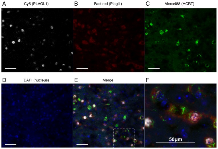
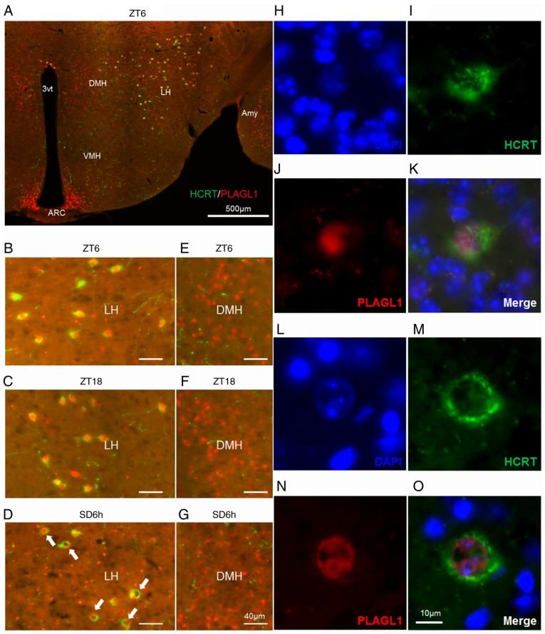
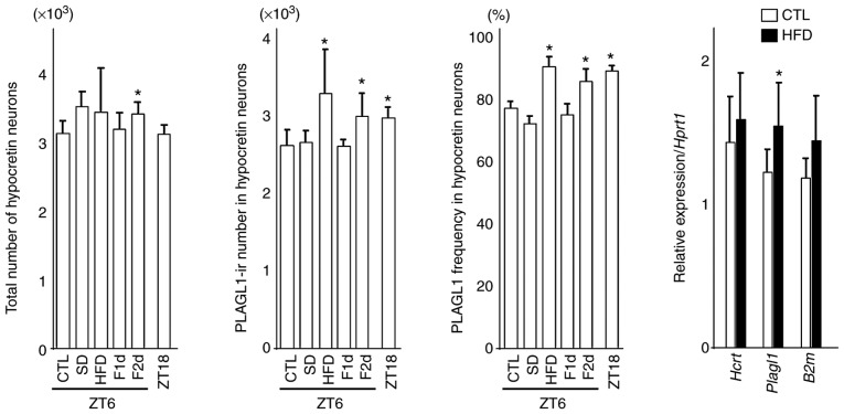
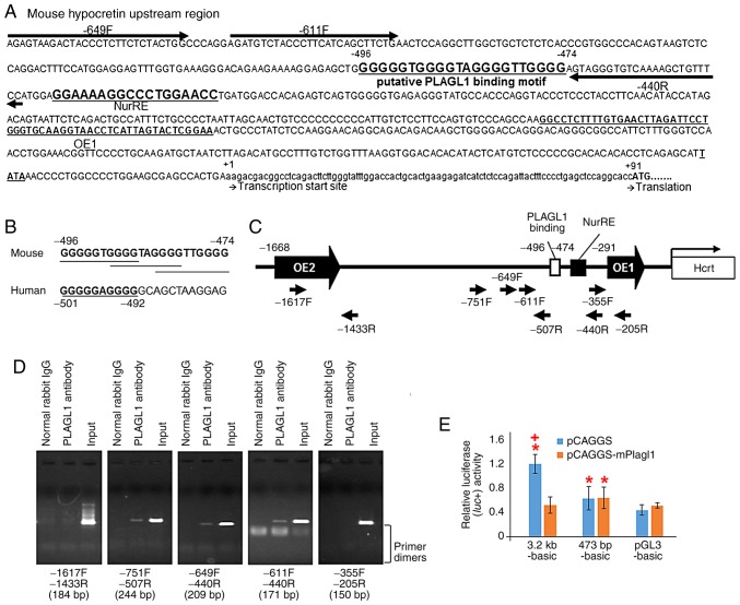
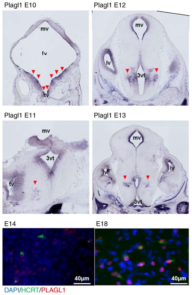
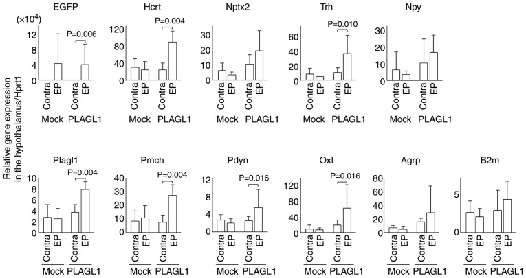
The hypocretin/orexin neuropeptide system coordinates the regulation of various physiological processes. Our previous study reported that a reduction in the expression of pleomorphic adenoma gene‑like 1 (Plagl1), which encodes a C2H2 zinc‑finger transcription factor, occurs in hypocretin neuron‑ablated transgenic mice, suggesting that PLAGL1 is co‑expressed in hypocretin neurons and regulates hypocretin transcription. The present study examined whether canonical prepro‑hypocretin transcription is functionally modulated by PLAGL1. Double immunostaining indicated that the majority of hypocretin neurons were positive for PLAGL1 immunoreactivity in the nucleus. Notably, PLAGL1 immunoreactivity in hypocretin neurons was altered in response to several conditions affecting hypocretin function. An uneven localization of PLAGL1 was detected in the nuclei of hypocretin neurons following sleep deprivation. Chromatin immunoprecipitation revealed that endogenous PLAGL1 may bind to a putative PLAGL1‑binding site in the proximal region of the hypocretin gene, in the murine hypothalamus. In addition, electroporation of the PLAGL1 expression vector into the fetal hypothalamus promoted hypothalamic hypocretin transcription. These results suggested that PLAGL1 may regulate hypothalamic hypocretin transcription.


Figures

Figure 1
Figure 1
Double immunohistochemical staining and RNAscope in the murine lateral hypothalamic area at ZT6. (A) PLAGL1 was visualized by Cy5 (white), (B) Plagl1 was visualized by Fast Red (red), (C) HCRT was visualized by Alexa Fluor® 488 (green), (D) and DAPI labeled the nuclei (blue). (E) Merged image of A-D; (F) expanded views of the square field in E. Scale bar, 50 µm. ZT, Zeitgeber time; HCRT, hypocretin; PLAGL1, pleomorphic adenoma gene-like 1.
Figure 2
Figure 2
Localization of PLAGL1 in murine hypocretin neurons under several conditions. (A-G) Murine HCRT was visualized by staining with Alexa Fluor® 488 (green) and murine PLAGL1 was visualized by staining with Alexa Fluor® 594 (red). (A) Murine hypothalamic sections at ZT6. The lateral hypothalamic area under (B) ZT6, (C) ZT18 and (D) SD6h conditions, with a relatively specific and uneven distribution of PLAGL1 (arrows). The dorsomedial hypothalamic nucleus under (E) ZT6, (F) ZT18 and (G) SD6h conditions. (H) DAPI-labeled nuclei at ZT6. (I) Alexa Fluor®488-labeled murine HCRT at ZT6. (J) Alexa Fluor® 594-labeled murine PLAGL1 at ZT6. (K) Merged image of (H, blue), (I, green) and (J, red) immunofluorescence at ZT6. (L) DAPI-labeled nuclei at SD6h. (M) Alexa Fluor®488-labeled murine HCRT at SD6h. (N) Alexa Fluor®594-labeled murine PLAGL1 at SD6h. (O) Merged image of (L, blue), (M, green), and (N, red) immunofluorescence at SD6h. Scale bar, (A) 500 µm, (B-G) 40 µm, (O) 10 µm. (H-O) These images are displayed at the same magnification. 3vt, third ventricle; Amy, amygdala; ARC, arcuate nucleus; DMH, dorsomedial hypothalamic nucleus; HCRT, hypocretin; LH, lateral hypothalamus; PLAGL1, pleomorphic adenoma gene-like 1; SD6h, 6 h of sleep deprivation; VMH, ventromedial nucleus of the hypothalamus; ZT, Zeitgeber time.
Figure 3
Figure 3
Number of hypocretin neurons, PLAGL1-ir number in hypocretin neurons, frequency of PLAGL1 and relative gene expression in hypocretin neurons. Differences in the count of hypocretin neurons, PLAGL1 counts and PLAGL1 immunopositivity were compared using one-way analysis of variance followed by Steel's post hoc test. *P<0.05 vs. the control (CTL) group (ZT6). Four or five animals from each group were used for each immunohistochemical analysis. The differences in relative gene expression were compared using two-tailed Student's t-tests for hypocretin and Plagl1 expression, and two-tailed Welch's t-test for B2m. Eight animals from each group were used for gene expression analysis. B2m, β-2-microglobulin; CTL, control (mice sacrificed at ZT6); F1d, 1 day of fasting; F2d, 2 days of fasting; Hcrt, hypocretin; HFD, high-fat diet; ir, immunoreactive; Plagl1, pleomorphic adenoma gene-like 1; SD, 6 h of sleep deprivation; ZT, Zeitgeber time; ZT18, mice sacrificed at ZT18. Bars indicate means, and error bars represent standard deviations.
Figure 4
Figure 4
Schematic representation of the murine prepro-hypocretin gene regulatory region, putative PLAGL1 binding sites in mice and humans, ChIP-PCR and luciferase reporter assay. (A) Sequence of the murine prepro-hypocretin gene regulatory region. Highly conserved region orexin regulatory element 1, which has been previously reported (2,3). The first residues of the transcription start site and the translation start site are marked as +1 and +91, respectively. NurRE (8), the putative PLAGL1 binding site and primers for ChIP-PCR analysis are shown. (B) Putative PLAGL1 binding sites in mouse and human promoter sequences are shown in bold and are underlined. (C) Schematic representation of the murine prepro-hypocretin gene regulatory region, and the position of ChIP-PCR primers. (D) ChIP-PCR analysis of endogenous PLAGL1 binding using ChIP-PCR primers. The length of each PCR product is represented at the bottom of each image. (E) Transcriptional activities of the human prepro-hypocretin promoter and murine deletion mutants in NIH3T3 cells. *P<0.05 vs. pGL3-basic with pCAGGS vector. +P<0.05 vs. each reporter with pCAGGS-mPlagl1. ChIP, chromatin immunoprecipitation; IgG, immunoglobulin G; NurRE, nuclear receptor response element; PLAGL1, pleomorphic adenoma gene-like 1; PCR, polymerase chain reaction.
Figure 5
Figure 5
Plagl1 expression in embryonic murine brains and localization of PLAGL1 in hypocretin neurons of the embryonic murine hypothalamus. The Plagl1 RNA probe detected Plagl1 expression in periventricular zones and the hypothalamus at E10-E13 by in situ hybridization (red arrowheads) (magnification, x20). Murine HCRT is visualized by staining with Alexa Fluor®488 (green) and murine PLAGL1 is visualized by staining with Alexa Fluor® 594 (red) at E14 and E18. 3vt, third ventricle; E, embryonic day; fv, forebrain vesicle; HCRT, hypocretin; mv, mesencephalic vesicle; PLAGL1, pleomorphic adenoma gene-like 1; tv, telencephalic vesicle.
Figure 6
Figure 6
Relative expression levels of hypothalamic genes following in utero electroporation. In utero electroporation was performed with pCAGGS-EGFP and pCAGGS-mPlagl1 (PLAGL1 group, n=6) to evaluate the effect of PLAGL1 on Hcrt transcription. Control mice were electroporated with pCAGGS-EGFP and pCAGGS (mock group, n=4). Animals were harvested at P0 stage. Following total RNA extraction from the hypothalamus and cDNA synthesis, quantitative polymerase chain reaction was performed. The relative expression levels of each gene were normalized to the relative expression levels of Hprt1. Alterations in transcription levels between the electroporated and contra sides in each group were compared using the Mann-Whitney U-test. Data are presented as the means ± standard deviation. Agrp, agouti related neuropeptide; B2m, β-2-microglobulin; Contra, contralateral side (no electroporation); EGFP, enhanced green fluorescent protein; EP, electroporated side; Hcrt, hypocretin; Hprt1, hypoxanthine phosphoribosyltransferase 1; Nptx2, neuronal pentaxin 2; Npy, neuropeptide Y; Oxt, oxytocin; Pdyn, prodynorphin; Pmch, pro-melanin-concentrating hormone; Trh, thyrotropin-releasing hormone.

Similar articles

Cited by

References

    1. Sakurai T. The role of orexin in motivated behaviours. Nat Rev Neurosci. 2014;15:719–731. doi: 10.1038/nrn3837. - DOI - PubMed
    1. Sakurai T, Moriguchi T, Furuya K, Kajiwara N, Nakamura T, Yanagisawa M, Goto K. Structure and function of human prepro-orexin gene. J Biol Chem. 1999;274:17771–17776. doi: 10.1074/jbc.274.25.17771. - DOI - PubMed
    1. Moriguchi T, Sakurai T, Takahashi S, Goto K, Yamamoto M. The human prepro-orexin gene regulatory region that activates gene expression in the lateral region and represses it in the medial regions of the hypothalamus. J Biol Chem. 2002;277:16985–16992. doi: 10.1074/jbc.M107962200. - DOI - PubMed
    1. Amiot C, Brischoux F, Colard C, La Roche A, Fellmann D, Risold PY. Hypocretin/orexin-containing neurons are produced in one sharp peak in the developing ventral diencephalon. Eur J Neurosci. 2005;22:531–534. doi: 10.1111/j.1460-9568.2005.04224.x. - DOI - PubMed
    1. Tanaka S. Transcriptional regulation of the hypocretin/orexin gene. Vitam Horm. 2012;89:75–90. doi: 10.1016/B978-0-12-394623-2.00005-6. - DOI - PubMed