. 2019 May;19(5):3497-3504.
doi: 10.3892/mmr.2019.10050. Epub 2019 Mar 18.

Enhanced sensitivity of capture IgE‑ELISA based on a recombinant Der f 1/2 fusion protein for the detection of IgE antibodies targeting house dust mite allergens

Affiliations

Enhanced sensitivity of capture IgE‑ELISA based on a recombinant Der f 1/2 fusion protein for the detection of IgE antibodies targeting house dust mite allergens

Zhen Zhang et al. Mol Med Rep. 2019 May.

Abstract

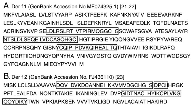
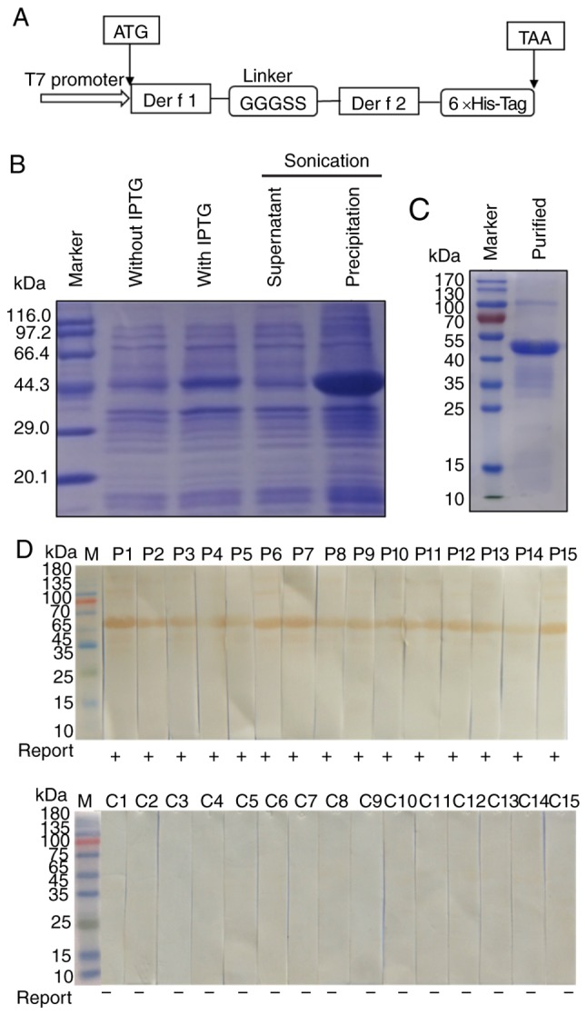
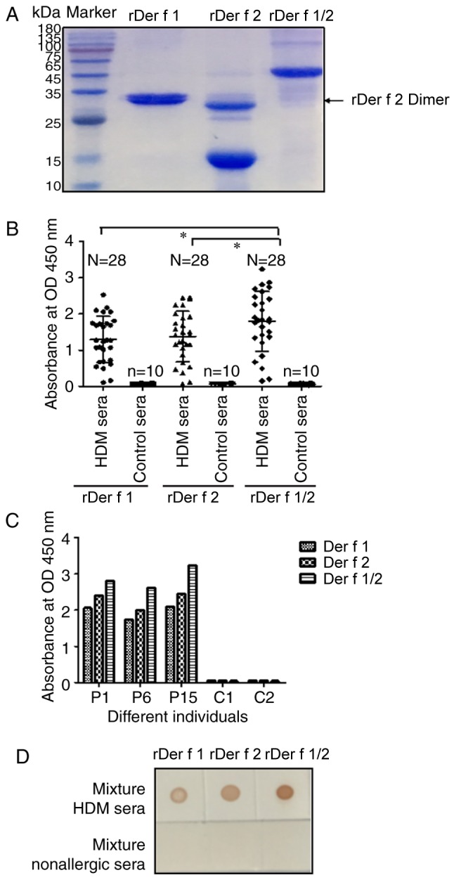
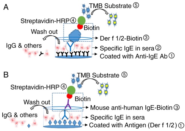
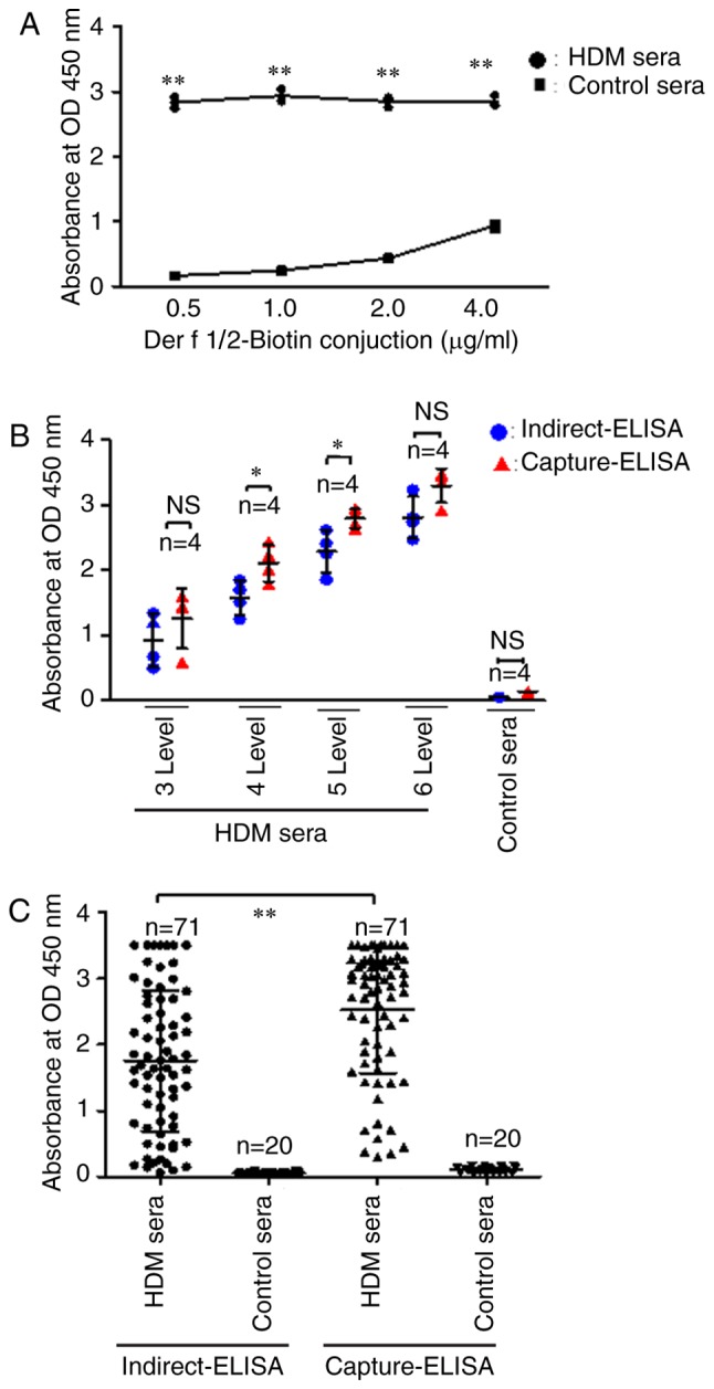
The detection of allergen‑specific immunoglobulin (Ig)E is an important method for the diagnosis of IgE‑mediated allergic diseases. The sensitivity of the indirect IgE‑ELISA method against allergen extracts is limited by interference from high IgG titers and low quantities of effectual allergen components in extracts. To overcome these limitations, a novel capture IgE‑ELISA based on a recombinant Der f 1/Der f 2 fusion protein (rDer f 1/2) was developed to enhance the sensitivity to IgEs that bind allergens from the house dust mite (HDM) species Dermatophagoides farina. pET28‑Der f 1/2 was constructed and expressed in Escherichia coli BL21 (DE3) pLysS. The purified fusion protein was evaluated by IgE western blotting, IgE dot blotting and indirect IgE‑ELISA. Capture‑ELISA was performed by coating wells with omalizumab and incubating in series with sera, biotinylated Der f 1/2, horseradish peroxidase‑conjugated streptavidin and 3,3,5,5‑tetramethylbenzidine. The relative sensitivities of indirect‑ELISA and capture‑ELISA for HDM allergen‑specific IgE binding were determined; sera from non‑allergic individuals were used as the control group. rDer f 1/2 was expressed in the form of inclusion bodies comprising refolded protein, which were then purified. It exhibited increased IgE‑specific binding (24/28, 85.8%) than rDer f 1 (21/28, 75.0%) or rDer f 2 (22/28, 78.6%) with HDM‑allergic sera. Furthermore, in a random sample of HDM‑allergic sera (n=71), capture‑ELISA (71/71, 100%) was more sensitive than indirect‑ELISA (68/71, 95.8%) for the detection of HDM‑specific IgEs (P<0.01), indicating that this novel method may be useful for the diagnosis of HDM allergy.


Figures

Figure 1.
Figure 1.
Reported linear B cell epitopes located in Der f 1 and Der f 2. Boxes indicate the locations of epitopes in (A) Der f 1 (GenBank accession no. MF074325.1) and (B) Der f 2 (GenBank accession no. FJ436110). The epitope peptide sequences for Der f 1 were 113LDLRSLRTVTPIRMQGGCG131, 151NTSLDLSEQELVDCASQHGC170 and 216CQIYPPDVKQIREALTQ232, whereas those for Der f 2 were 18DQVDVKDCANNEIKKVMVDGCHGS41, 42DPCII46 and 74GIDTNACHYIKCPLVKGQQYDIKY97. Der f, allergen from Dermatophagoides farina.
Figure 2.
Figure 2.
Expression, purification and IgE binding activity of rDer f 1/2. (A) Schematic diagram of the Der f 1/2 fusion protein construct. (B) SDS-PAGE of rDer f 1/2 following expression in Escherichia coli BL21(DE3) pLysS. (C) SDS-PAGE of rDer f 1/2 following purification by nickel affinity chromatography. Western blotting was conducted to determine the binding of rDer f 1/2 to specific IgE in the sera from (D) 15 house dust mite-allergic patients or (C) 15 non-allergic controls. 6×His-Tag, hexahistidine tag; Der f, allergen from Dermatophagoides farina; Ig, immunoglobulin; IPTG, isopropyl-β-D-thiogalactopyranoside; r, recombinant.
Figure 3.
Figure 3.
Analysis of the binding of specific IgE to rDer f 1, rDer f 2 and rDer f 1/2 in HDM sera. (A) SDS-PAGE of purified recombinant proteins. (B) Levels of binding of rDer f 1, rDer f 2 and rDer f 1/2 to IgE in 28 HDM and 10 control sera as determined by indirect ELISA. (C) Binding of rDer f 1, rDer f 2, and rDer f 1/2 to specific IgE in sera from individuals with HDM allergies (P1, P6 and P15) and non-allergic controls (C1 and C2) as determined by indirect ELISA. (D) Recombinant proteins were dotted onto nitrocellulose strips and incubated with mixed sera from 3 HDM-allergic patients or 3 non-allergic individuals. Data are presented as the mean ± standard error of the mean. *P<0.05. Control sera, sera from non-allergic control individuals; Der f, allergen from Dermatophagoides farina; HDM, house dust mite; HDM sera, sera from patients with HDM allergies; Ig, immunoglobulin; OD, optical density; r, recombinant.
Figure 4.
Figure 4.
Schematic diagrams of direct capture IgE-ELISA and indirect IgE-ELISA. (A) Direct capture and (B) indirect IgE-ELISAs for the detection of specific IgEs in HDM-allergic sera. For capture IgE-ELISA, 96-well plates were coated with anti-IgE Ab (omalizumab), blocked and incubated with HDM-allergic sera containing HDM-specific IgE, prior to incubations with rDer f 1/2-Biotin and streptavidin-HRP. For indirect IgE-ELISA, plates were coated with rDer f 1/2, blocked and incubated with HDM-allergic sera, prior to incubations with biotinylated mouse anti-human IgE and streptavidin-HRP. For the two methods, TMB substrate was subsequently added; the reaction was terminated following the addition of 2 M H2SO4. Ab, antibody; Der f, allergen from Dermatophagoides farina; Der f 1/2-biotin, biotinylated Der f 1/2 protein; HDM, house dust mite; HDM-allergic sera, sera from patients with HDM allergies; HRP, horseradish peroxidase; Ig, immunoglobulin; r, recombinant; TMB, 3,3′,5,5′-tetramethylbenzidine.
Figure 5.
Figure 5.
Capture-ELISA detection of specific IgEs in HDM sera. (A) Identification of optimal Der f1/2-biotin conjugate concentration for capture ELISA. (B) Sensitivity of indirect and capture ELISAs in the detection of allergen-specific IgE in HDM serum samples with varying levels of IgE as determined by ImmunoCAP. IgE levels were defined as follows: Level 3, 3.5–17.5 IU/ml; level 4, 17.5–50.0 IU/ml; level 5, 50.0–100 IU/ml and level 6, >100 IU/ml. (C) Sensitivity of indirect and capture ELISAs in the detection of allergen-specific IgE in a random cohort of HDM serum samples (n=71). Data are presented as the mean ± standard error of the mean. *P<0.05, **P<0.01. Control sera, sera from non-allergic control individuals; Der f, allergen from Dermatophagoides farina; Der f 1/2-biotin, biotinylated Der f 1/2 protein; HDM, house dust mite; HDM sera, sera from patients with HDM allergies; Ig, immunoglobulin; OD, optical density.

Similar articles

Cited by

References

    1. Chan TF, Ji KM, Yim AK, Liu XY, Zhou JW, Li RQ, Yang KY, Li J, Li M, Law PT, et al. The draft genome, transcriptome, and microbiome of Dermatophagoides farinae reveal a broad spectrum of dust mite allergens. J Allergy Clin Immunol. 2015;135:539–548. doi: 10.1016/j.jaci.2014.09.031. - DOI - PubMed
    1. Elkady A. Allergy to Dermatophagoides pteronyssinus (Der p1) and Dermatophagoides farina (Der f1) in patients with atopic asthma. Int J Sci Res. 2015;4:1896–1902.
    1. An S, Chen L, Long C, Liu X, Xu X, Lu X, Rong M, Liu Z, Lai R. Dermatophagoides farinae allergens diversity identification by proteomics. Mol Cell Proteomics. 2013;12:1818–1828. doi: 10.1074/mcp.M112.027136. - DOI - PMC - PubMed
    1. Chang ML, Cui C, Liu YH, Pei LC, Shao B. Analysis of total immunoglobulin E and specific immunoglobulin E of 3,721 patients with allergic disease. Biomed Rep. 2015;3:573–577. doi: 10.3892/br.2015.455. - DOI - PMC - PubMed
    1. Williams P, Sewell WA, Pumphrey R, Read G, Jolles S. Clinical immunology review series: An approach to the use of the immunology laboratory in the diagnosis of clinical allergy. Clin Exp Immunol. 2010;153:10–18. doi: 10.1111/j.1365-2249.2008.03695.x. - DOI - PMC - PubMed

MeSH terms