. 2019 May;41(5):2729-2738.
doi: 10.3892/or.2019.7078. Epub 2019 Mar 19.

miR-363 acts as a tumor suppressor in osteosarcoma cells by inhibiting PDZD2

Affiliations

miR-363 acts as a tumor suppressor in osteosarcoma cells by inhibiting PDZD2

Fan He et al. Oncol Rep. 2019 May.

Abstract

PDZ domain containing 2 (PDZD2) is a multi-PDZ domain protein that promotes the proliferation of insulinoma cells, and is upregulated during prostate tumorigenesis. However, the function of PDZD2 in other cancers, including osteosarcoma (OS), remains unclear. Dysregulation of microRNAs (miRNAs) contributes to tumor initiation, proliferation and metastasis, via the regulation of their target genes. The present study investigated the functions of miR-363 and PDZD2 in MG-63 OS cells. The results revealed that MG-63 cells contained low levels of miR-363, and that overexpression of miR-363 in MG-63 cells significantly inhibited the vitality, proliferation, and colony formation ability of the cells, but promoted their apoptosis and G1/S arrest by regulating proliferating cell nuclear antigen (PCNA) and caspase-3 expression. Additionally, miR-363 impaired the migration and invasion of MG-63 cells by regulating the epithelial-mesenchymal transition (EMT) phenotype. Notably, a bioinformatics analysis and luciferase reporter assay indicated that PDZD2 was a direct target of miR-363. miR-363 overexpression reduced PDZD2 protein levels and knockdown of PDZD2 suppressed the colony formation, migration and invasion of MG-63 cells, but promoted their apoptosis by regulating expression of PCNA, caspase-3, and the EMT phenotype. In vivo studies further confirmed that miR-363 functioned as tumor suppressor, by inhibiting tumor growth, promoting cell apoptosis, and reducing PDZD2 and PCNA levels and the prevalence of the EMT phenotype in tumor tissues. The present data demonstrated that downregulation of the tumor suppressor miR-363 may be involved in the development of osteosarcoma via regulation of PDZD2.


Figures
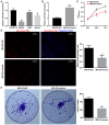
Figure 1.
Figure 1.
Restoration of miR-363 expression in OS cells inhibits cell proliferation in vitro. (A) Expression levels of miR-363 were determined in three OS cell lines (MG-63, HOS, and Saos2) and one human osteoblastic cell line (hFOB1.19) by reverse transcription-quantitative polymerase chain reaction. *P<0.05 and **P<0.01 compared with hFOB1.19 cells. (B) miR-363 mimics and negative control were efficiently transfected into MG-63 cells. (C) Cell viability (CCK-8 assay), (D) proliferation (EdU assay) and (E) colony formation were evaluated in the cells transfected with either miR-363 mimics or negative control. Data are presented as mean ± standard deviation. **P<0.01 and ***P<0.001 compared with NC group. OS, osteosarcoma; NC negative control; OD, optical density.
Figure 2.
Figure 2.
miR-363 overexpression in MG-63 cells induces apoptosis in vitro. (A) The apoptosis rate was determined by Hoechst staining (magnification, ×200) and (B) flow cytometry. (C) Cell cycle distribution was determined by PI staining and flow cytometry analysis. Data are presented as mean ± standard deviation. **P<0.01 compared with NC group. PI, propidium iodide; NC negative control; FITC, fluorescein isothiocyanate.
Figure 3.
Figure 3.
Restoration of miR-363 expression in osteosarcoma cells impairs their migration and invasion capabilities. (A) Migration of MG-63 cells was determined with the scratch wound assay. (B) Invasion of MG-63 cells was determined with the transwell assay (magnification, ×200). (C) Expression of EMT markers E-cadherin and vimentin was evaluated by immunofluorescence (magnification, ×200). (D) Western blot analysis of protein expression for the EMT markers as well as PZDZ2, PCNA, and cleaved caspase-3. Data are presented as mean ± standard deviation. **P<0.01 compared with NC group. EMT, epithelial-mesenchymal transition; NC negative control; PZDZ2, PDZ domain containing 2; PCNA, proliferating cell nuclear antigen.
Figure 4.
Figure 4.
miR-363 directly targets PDZD2 in osteosarcoma cells. (A) The 3′UTR of PDZD2 mRNA harbors a binding site for miR-363 (underlined sequence). (B) Overexpression of miR-363 reduced the levels of PDZD2 mRNA and (C) protein in MG-63 cells, as determined by reverse transcription-quantitative polymerase chain reaction and immunofluorescence analysis (magnification, ×200), respectively. (D) MG-63 cells were transfected with the full-length 3′UTR (wild-type or mutant) of PDZD2, and the luciferase reporter assay was used to confirm direct interaction. Data are presented as mean ± standard deviation. **P<0.01 and ***P<0.001 compared with NC group. PZDZ2, PDZ domain containing 2; UTR; untranslated region; NC negative control; WT, wild-type; MUT, mutant.
Figure 5.
Figure 5.
PDZD2 knockdown inhibits osteosarcoma cell growth and EMT. (A) Successful knockdown of PDZD2 was established by evaluating three separate siRNAs, and selecting the most efficient construct. (B) Cell apoptosis (magnification, ×200), (C) colony formation and (D) invasion (magnification, ×200) were evaluated in the MG-63 cells following PDZD2 knockdown. (E) Protein levels of PDZD2, EMT markers E-cadherin and vimentin, PCNA, and cleaved caspase-3 were determined by western blot analysis. Data are presented as mean ± standard deviation. **P<0.01 and ***P<0.001 compared with NC group. PZDZ2, PDZ domain containing 2; EMT, epithelial-mesenchymal transition; siRNA, small interfering RNA; PCNA, proliferating cell nuclear antigen; NC negative control.
Figure 6.
Figure 6.
mir-363 induces the regression of osteosarcoma tumors in vivo. MG-63 cells (~2×10⁶), transfected with miR-363 mimics or negative control, were subcutaneously injected into the rear flanks of nude mice (5 mice per group). (A) Images of the tumors at the end of the experiment. (B) Quantification of the tumor volumes (mm3) over time. (C) Expression levels of miR-363 and (D) PDZD2 mRNA in tumor tissues were analyzed by reverse transcription-quantitative polymerase chain reaction. (E) PDZD2 protein expression in tumor tissues was determined by immunohistochemistry (magnification, ×200). (F) The apoptosis rates in the tumor sections were estimated by TUNEL staining (magnification, ×200). (G) Protein expression levels of PDZD2, E-cadherin, vimentin, PCNA and cleaved caspase-3 expression were determined by western blot analysis. Data are presented as mean ± standard deviation. *P<0.05, **P<0.01 and ***P<0.001 compared with NC group. PZDZ2, PDZ domain containing 2; PCNA, proliferating cell nuclear antigen; NC negative control.

Similar articles

Cited by

References

    1. Geller DS, Gorlick R. Osteosarcoma: A review of diagnosis, management, and treatment strategies. Clin Adv Hematol Oncol. 2010;8:705–718. - PubMed
    1. Lamoureux F, Trichet V, Chipoy C, Blanchard F, Gouin F, Redini F. Recent advances in the management of osteosarcoma and forthcoming therapeutic strategies. Expert Rev Anticancer Ther. 2007;7:169–181. doi: 10.1586/14737140.7.2.169. - DOI - PubMed
    1. Gaspar N, Hawkins DS, Dirksen U, Lewis IJ, Ferrari S, Le Deley MC, Kovar H, Grimer R, Whelan J, Claude L, et al. Ewing sarcoma: Current management and future approaches through collaboration. J Clin Oncol. 2015;33:3036–3046. doi: 10.1200/JCO.2014.59.5256. - DOI - PubMed
    1. Misaghi A, Goldin A, Awad M, Kulidjian AA. Osteosarcoma: A comprehensive review. SICOT J. 2018;4:12. doi: 10.1051/sicotj/2017028. - DOI - PMC - PubMed
    1. Thomas MK, Tsang SW, Yeung ML, Leung PS, Yao KM. The roles of the PDZ-containing proteins bridge-1 and PDZD2 in the regulation of insulin production and pancreatic beta-cell mass. Curr Protein Pept Sci. 2009;10:30–36. doi: 10.2174/138920309787315248. - DOI - PubMed

MeSH terms