. 2019 Sep 15;200(6):742-750.
doi: 10.1164/rccm.201806-1178OC.

Driver Mutations in Normal Airway Epithelium Elucidate Spatiotemporal Resolution of Lung Cancer

Affiliations

Driver Mutations in Normal Airway Epithelium Elucidate Spatiotemporal Resolution of Lung Cancer

Humam Kadara et al. Am J Respir Crit Care Med. .

Abstract

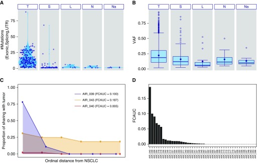
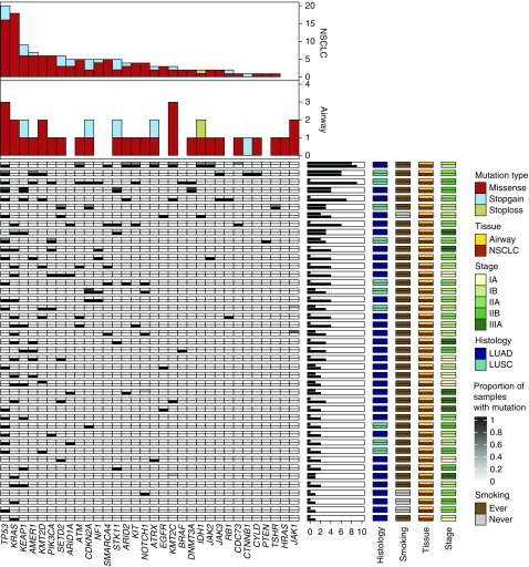
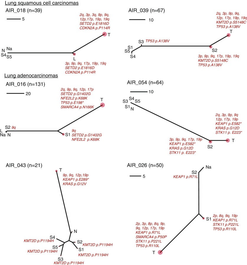
Rationale: Uninvolved normal-appearing airway epithelium has been shown to exhibit specific mutations characteristic of nearby non-small cell lung cancers (NSCLCs). Yet, its somatic mutational landscape in patients with early-stage NSCLC is unknown.Objectives: To comprehensively survey the somatic mutational architecture of the normal airway epithelium in patients with early-stage NSCLC.Methods: Multiregion normal airways, comprising tumor-adjacent small airways, tumor-distant large airways, nasal epithelium and uninvolved normal lung (collectively airway field), matched NSCLCs, and blood cells (n = 498) from 48 patients were interrogated for somatic single-nucleotide variants by deep-targeted DNA sequencing and for chromosomal allelic imbalance events by genome-wide genotype array profiling. Spatiotemporal relationships between the airway field and NSCLCs were assessed by phylogenetic analysis.Measurements and Main Results: Genomic airway field carcinogenesis was observed in 25 cases (52%). The airway field epithelium exhibited a total of 269 somatic mutations in most patients (n = 36) including key drivers that were shared with the NSCLCs. Allele frequencies of these acquired variants were overall higher in NSCLCs. Integrative analysis of single-nucleotide variants and allelic imbalance events revealed driver genes with shared "two-hit" alterations in the airway field (e.g., TP53, KRAS, KEAP1, STK11, and CDKN2A) and those with single hits progressing to two in the NSCLCs (e.g., PIK3CA and NOTCH1).Conclusions: Tumor-adjacent and tumor-distant normal-appearing airway epithelia exhibit somatic driver alterations that undergo selection-driven clonal expansion in NSCLC. These events offer spatiotemporal insights into the development of NSCLC and, thus, potential targets for early treatment.

Keywords: allelic imbalance; cancerization field; deep targeted sequencing; early-stage non–small cell lung cancer; normal airway.


Figures

Figure 1.
Figure 1.
Somatic mutation profiles in the genomic cancerization field of the normal airway in early-stage non–small cell lung cancer (NSCLC). Deep DNA sequencing and SNP array profiling analyses of 450 multiregion normal-appearing airway epithelia and NSCLCs, along with 48 germline samples, from 48 patients with early-stage NSCLC, were performed as described in the Methods and online supplement. (A) The total number of somatic single-nucleotide variants (SNVs) across the airway field comprising multiregion samples from tumor-adjacent small airways (S), distant large airways (L), nasal epithelium (Na), and uninvolved normal lung tissue (N) and their matched NSCLCs (T) are depicted. Each point represents a single sample and plots within each sample type show somatic SNV burden distributions. (B) Box plots demonstrate variant allele frequency distributions of the identified SNVs across the different types of samples. (C) Genomic airway field cancerization was quantified based on shared SNV and allelic imbalance profiles as described in the Methods and online supplement sections and summarized as field cancerization area under the curve (FCAUC: 0, lack of airway cancerization evidence and no sharing of alterations in the airway field with the tumor; 1, complete sharing of alterations between all airway field samples and matched NSCLCs). Shown here are three representative cases with relatively varied FCAUCs. The x-axis denotes an ordinal distance of airway field tissues from its matched NSCLC (0–1), and y-axis denotes the proportion of shared aberrations with the matched NSCLC (0 indicates no shared events, to 1 for complete sharing). The area under the curve (i.e., FCAUC) for each of the three cases are shaded. (D) A bar plot of FCAUCs for each patient profiled. UTR = untranslated region; VAF = variant allele frequency.
Figure 2.
Figure 2.
Landscape of somatic driver variants in the non–small cell lung cancer (NSCLC)-adjacent and NSCLC-distant normal airway epithelium. Deep DNA sequencing of a cancer gene panel (n = 409) and identification of somatic nonsynonymous (e.g., missense, nonsense, and stoploss) variants in all airway field and matched NSCLCs was performed as described in the Methods section. Mutated genes previously implicated as drivers in NSCLC or other malignancies (–19) are shown for the airway field and tumor samples. Columns denote genes and rows represent individual patients. Each patient is denoted by a row with the airway field presented on the top half of the cell and the matched NSCLC in the bottom half. Mutated genes are color coded based on the proportion of airway samples carrying a variant within the gene (proportion range 0–1; white to black; right panel) and presence in the matched NSCLC (white, absent; black, present; right panel). The number of patients with the indicated driver mutated genes in the airway field and NSCLC are shown as bar plots (top panels). Annotations for stage, histology, smoking, and tissue type (airway and NSCLCs) for all patients are shown in the right panels. Patients were ordered, top to bottom, based on airway field and NSCLC somatic mutation burdens (middle horizontal barplots). LUAD = lung adenocarcinoma; LUSC = lung squamous cell carcinoma.
Figure 3.
Figure 3.
Somatic “two-hit” aberrations in the adjacent and distant normal airway epithelium of patients with early-stage non–small cell lung cancer (NSCLC). Data from deep DNA sequencing and SNP array profiling were integrated to identify NSCLC-associated drivers that comprised either somatic single-nucleotide variants (SNVs) or allelic imbalance (AI) and genes with two-hit aberrations (both SNVs and AI) in the airway field and NSCLC samples. Columns and rows represent patients and NSCLC-associated driver genes, respectively. Each column denotes a patient with the left half of the cell corresponding to the airway field (gray) and right half (black) to its matched NSCLC. Genes with two-hit aberrations are depicted in red and genes comprising either single SNVs or AI events are depicted in orange and yellow, respectively. The detected AI events were annotated as gain (brown), loss (blue), copy-neutral loss-of-heterozygosity (cn-LOH, green), and undeterminable (gray) events. AI events exhibiting intratumoral heterogeneity within multiregion tumor samples (e.g., one biopsy with a cn-LOH and another biopsy within the same tumor showing a copy gain for the same chromosomal region: cn-LOH, gain) are annotated separately. Stacked bars showing the distribution of different types of AI events for each gene are shown in the right panels for both the airway fields and NSCLCs. NSCLC-associated driver genes are ordered top to bottom based on overall two-hit and single-hit patterns in the airway field and NSCLC; and the cases (columns) are ordered left to right based on overall burden of somatic hits across these genes. LUAD = lung adenocarcinoma; LUSC = lung squamous cell carcinoma.
Figure 4.
Figure 4.
Molecular spatial and temporal relationships between the normal airway cancerization field and early-stage non–small cell lung cancer (NSCLC). For every patient, the single-nucleotide variants and allelic imbalances detected (n) across airway field and NSCLC tissues were integrated to generate unrooted neighbor-joining phylogenetic trees to study intrapatient multiregion samples as described in the Methods section. Six cases (two LUSC and four LUAD) previously identified by an independent statistical analysis of field cancerization (Figure 1D) are shown. The phylogenetic trees were annotated with mutations in known cancer-associated genes and large chromosomal aberrations previously implicated in NSCLC pathogenesis. Each tree is accompanied by a scale to denote the number of mutations. The relative somatic burden for each tissue in a tree is denoted by a correspondingly sized red circle. The distances among the multiple points of a tree correspond to the extent of shared and disparate mutational events among samples of a patient. The tree topologies for cases with evidence for genomic field cancerization differ from a typically straight line that would be expected if only NSCLCs presented mutations (see online supplement). Phylogenetic trees for the remaining cases are provided in the online supplement. L = distant large airways; LUAD = lung adenocarcinoma; LUSC = lung squamous cell carcinoma; N = uninvolved normal lung tissue; Na = nasal epithelium; S = tumor-adjacent small airways; T = matched NSCLC.

Comment in

  • Sequencing Lung Cancer's Sequence.
    Watanabe H, Powell CA. Watanabe H, et al. Am J Respir Crit Care Med. 2019 Sep 15;200(6):657-659. doi: 10.1164/rccm.201904-0837ED. Am J Respir Crit Care Med. 2019. PMID: 31059279 Free PMC article. No abstract available.

References

    1. Slaughter DP, Southwick HW, Smejkal W. Field cancerization in oral stratified squamous epithelium; clinical implications of multicentric origin. Cancer. 1953;6:963–968. - PubMed
    1. Curtius K, Wright NA, Graham TA. An evolutionary perspective on field cancerization. Nat Rev Cancer. 2018;18:19–32. - PubMed
    1. Kadara H, Wistuba II. Field cancerization in non-small cell lung cancer: implications in disease pathogenesis. Proc Am Thorac Soc. 2012;9:38–42. - PMC - PubMed
    1. Braakhuis BJ, Tabor MP, Kummer JA, Leemans CR, Brakenhoff RH. A genetic explanation of Slaughter’s concept of field cancerization: evidence and clinical implications. Cancer Res. 2003;63:1727–1730. - PubMed
    1. Siegel RL, Miller KD, Jemal A. Cancer statistics, 2018. CA Cancer J Clin. 2018;68:7–30. - PubMed

Publication types