. 2019 Mar 21;17(3):e2006540.
doi: 10.1371/journal.pbio.2006540. eCollection 2019 Mar.

Comprehensive profiling of the STE20 kinase family defines features essential for selective substrate targeting and signaling output

Affiliations

Comprehensive profiling of the STE20 kinase family defines features essential for selective substrate targeting and signaling output

Chad J Miller et al. PLoS Biol. .

Abstract

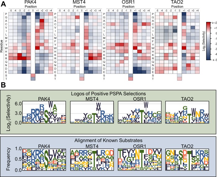
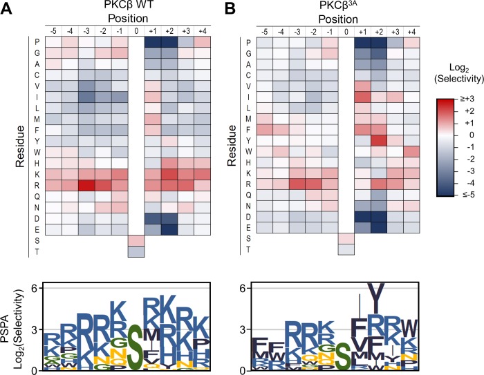
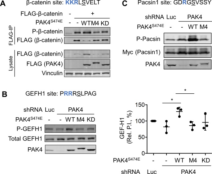
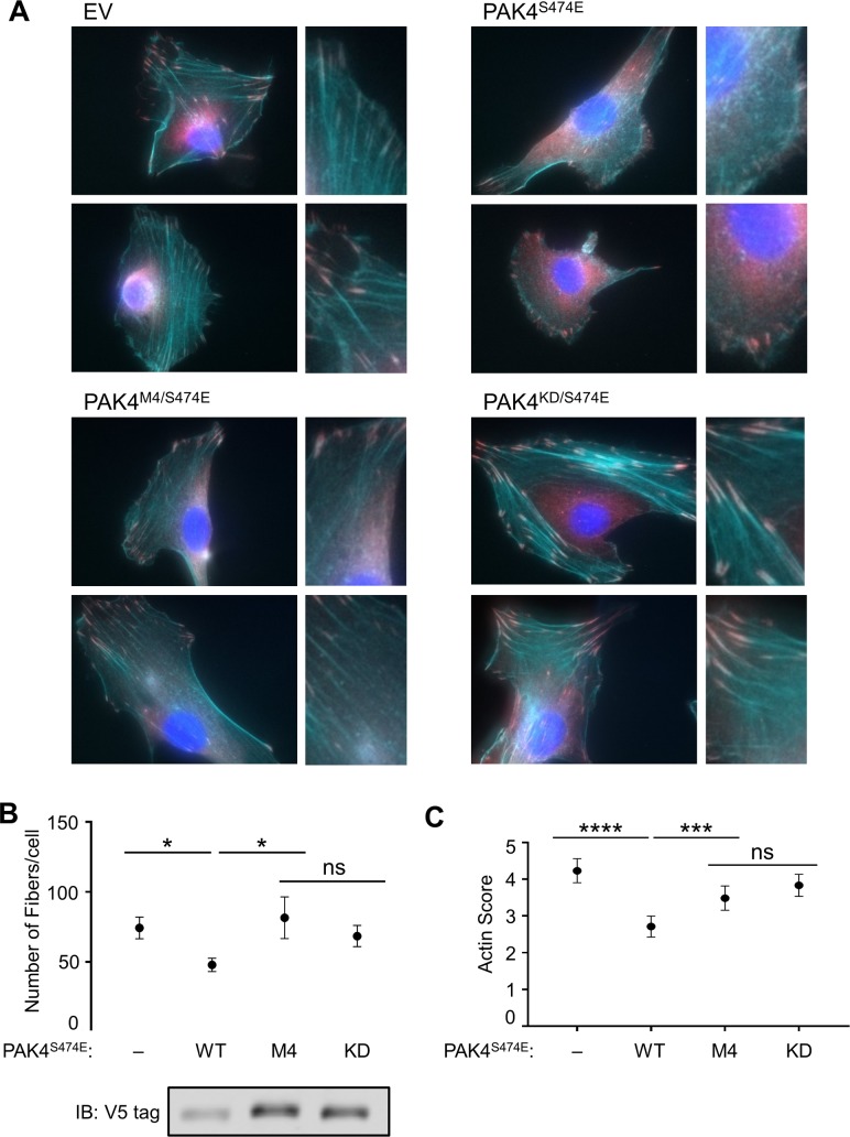
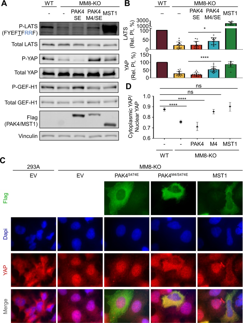
Specificity within protein kinase signaling cascades is determined by direct and indirect interactions between kinases and their substrates. While the impact of localization and recruitment on kinase-substrate targeting can be readily assessed, evaluating the relative importance of direct phosphorylation site interactions remains challenging. In this study, we examine the STE20 family of protein serine-threonine kinases to investigate basic mechanisms of substrate targeting. We used peptide arrays to define the phosphorylation site specificity for the majority of STE20 kinases and categorized them into four distinct groups. Using structure-guided mutagenesis, we identified key specificity-determining residues within the kinase catalytic cleft, including an unappreciated role for the kinase β3-αC loop region in controlling specificity. Exchanging key residues between the STE20 kinases p21-activated kinase 4 (PAK4) and Mammalian sterile 20 kinase 4 (MST4) largely interconverted their phosphorylation site preferences. In cells, a reprogrammed PAK4 mutant, engineered to recognize MST substrates, failed to phosphorylate PAK4 substrates or to mediate remodeling of the actin cytoskeleton. In contrast, this mutant could rescue signaling through the Hippo pathway in cells lacking multiple MST kinases. These observations formally demonstrate the importance of catalytic site specificity for directing protein kinase signal transduction pathways. Our findings further suggest that phosphorylation site specificity is both necessary and sufficient to mediate distinct signaling outputs of STE20 kinases and imply broad applicability to other kinase signaling systems.


Conflict of interest statement

The authors have declared that no competing interests exist.

Figures

Fig 1
Fig 1. PSPA analysis showing representatives of the four major STE20 specificity groups.
(A) Quantified PSPA spot intensities were normalized to an average value of 1 at each position within the peptide. Log2 transformed data are depicted as heat maps showing positively and negatively selected residues by position. Data are the mean of at least two separate experiments. Numerical values are provided in S1 Data. (B) Top: Positively selected residues from PSPA analysis for the same kinases as in panel A are shown as sequence logos. Bottom: Sequence logos based on alignments of all known phosphorylation sites mapped on protein substrates for the indicated kinase. Substrate phosphorylation sites were collected from the PhosphoSitePlus database, with the exception of TAO2 substrate sites that were identified in [43]. Logos were generated with enoLOGOS [44]. MST, Mammalian sterile 20 kinase; OSR1, Oxidative stress-responsive 1; PAK, p21-activated kinase; PSPA, positional scanning peptide array; TAO2, thousand and one amino acid kinase 2
Fig 2
Fig 2. Phosphorylation kinetics of peptide substrates by STE20 kinases.
(A) Michaelis–Menten curve for MST4 phosphorylation of MSTtide. Individual data points from three separate experiments are shown. (B) Initial rates of phosphorylation by MST4 of a series of peptides with the indicated sequences (n ≥ 3, bars show mean ± SD) shown relative to MSTtide (top) phosphorylation. Data for PAKtide are at bottom. (C) Relative initial rates of phosphorylation of MSTtide and PAKtide by a series of STE20 kinases (n = 3, error bars are 95% CI). Kinases with bars that do not cross the y-axis have a statistically significant preference for one consensus peptide over the other (p = 0.05). Source data for all panels are provided in S2 Data. GCK, germinal center kinase; HGK, HPK/GCK-like kinase; HPK1, Hematopoietic progenitor kinase 1; KHS, Kinase homologous to SPS1/STE20; LOK, Lymphocyte-oriented kinase; MINK, Misshapen-like kinase 1; MST, Mammalian sterile 20 kinase; MYO, myosin; OSR1, Oxidative stress-responsive 1; PAK, p21-activated kinase; SLK, STE20-like kinase; TAO, thousand and one amino acid kinase; YSK1, Yeast Sps1/Ste20-related Kinase 1
Fig 3
Fig 3. Overview of putative specificity-determining residues in the STE kinase family.
(A) Regions of the kinase proximal to the −2 (KxxN motif), 0 (DFG+1), and +2/+3 (β3–αC loop region) residues are highlighted on the x-ray crystal structure of PAK4 in complex with a peptide substrate (PDB: 2Q0N). (B) Dendrogram of the STE20 kinase family showing putative specificity-determining regions and corresponding residues selected by PSPA analysis. The alignment of the β3–αC loop region was made using data from available x-ray crystal structures and predictions from PSIPRED v3.3 [49, 50]. GCK, germinal center kinase; HGK, HPK/GCK-like kinase; HPK, Hematopoietic progenitor kinase 1; KHS, Kinase homologous to SPS1/STE20; LOK, Lymphocyte-oriented kinase; MINK, Misshapen-like kinase 1; MST, Mammalian sterile 20 kinase; MYO, myosin; NRK, NIK-related protein kinase; OSR1, Oxidative stress-responsive 1; PAK, p21-activated kinase; PDB, Protein Data Bank; PSPA, positional scanning peptide array; SLK, STE20-like kinase; SPAK, STE20/SPS1-related proline-alanine–rich kinase; TAO, thousand and one amino acid; TNIK, Traf2 and NCK-interacting protein kinase; YSK1, Yeast Sps1/Ste20-related Kinase 1
Fig 4
Fig 4. Mutation of specificity-determining residues exchanges substrate specificity between PAK4 and MST4.
Mutants harbor residues found at the indicated positions in the other kinase as described in the main text. The combination mutants (PAK4M4 and MST4P4) include KxxN, DFG+1 and β3–αC loop region mutants (and S445N for PAK4M4). (A) Logos show positively selected residues from PSPA analysis of the indicated mutant kinases. Data were processed and visualized as in Fig 1. Bar graphs show peptide kinase assays with the indicated pairs of peptide substrates (n = 3, error bars indicate SD, rate units are nM/min/nM kinase). (B) Autoradiograph shows in vitro phosphorylation of full-length β-catenin variants with the indicated sequences surrounding Ser675 by WT PAK4. SA and TA are the corresponding S675A or T675A mutants. All β-catenin constructs included two additional mutations to remove minor sites of phosphorylation (S552A/T556A). (C) The graph shows the level of WT PAK4 phosphorylation of β-catenin variants used in (B) over time under pre-steady–state conditions. Numerical data for (A) and (C) are in S2 Data. MST, Mammalian sterile 20 kinase; PAK, p21-activated kinase; PSPA, positional scanning peptide array; WT, wild type.
Fig 5
Fig 5. Acidic residues in the β3–αC loop region promote selection of basic residues by PKCβ.
PSPA analysis shown as heat maps and sequence logos (prepared as in Fig 1) for WT (A) and the β3–αC loop mutant (B) of PKCβ. Numerical data are provided in S1 Data. PKC, protein kinase C; PSPA, positional scanning peptide array; WT, wild type.
Fig 6
Fig 6. Catalytic site interactions are required for PAK4 to target protein substrates in cells.
(A) HEK293A cells were co-transfected with plasmids expressing FLAG-epitope–tagged β-catenin and PAK4S474E with the additional mutations as indicated. Following FLAG immunoprecipitation from cell lysates, equal amounts of purified β-catenin were subjected to immunoblotting to detect phosphorylation at Ser675. (B) Panc1 cells expressing a doxycycline-inducible shRNA directed to PAK4 or a nontargeting control shRNA were transfected with plasmids expressing the indicated forms of PAK4S474E. Phosphorylation of endogenous GEF-H1 at Ser886 was assessed by immunoblotting with a phosphospecific antibody. The phosphorylation index (ratio of phospho-GEF-H1 to total GEF-H1) was quantified and normalized to the empty vector control (n = 3, error bars indicate SD). Empty vector, PAK4S474E, and PAK4M4/S474E signals were compared with unpaired t tests (*p < 0.05). Numerical data are provided in S2 Data. (C) The Panc1 cell lines used in (B) were co-transfected with plasmids expressing Myc-epitope–tagged Pacsin1 and the indicated PAK4S474E mutants, and Pacsin1 phosphorylation at Ser346 was analyzed by immunoblotting. GEF-H1, Rho guanine nucleotide exchange factor H1; HEK, human embryonic kidney; IP, immunoprecipitation; KD, kinase-inactive mutant; Pacsin1, Protein kinase C and casein kinase substrate in neurons protein 1; PAK, p21-activated kinase; P.I., phosphorylation index; P-, phospho-; shRNA, short hairpin RNA; WT, wild type.
Fig 7
Fig 7. Phosphorylation site specificity is required for actin disassembly by PAK4.
(A) NIH-3T3 cells stably expressing the indicated PAK4 mutants were imaged following staining with phalloidin (cyan), α-vinculin (magenta), and DAPI (blue). (B) The number of actin fibers per cell were identified using SFEX software [54] to analyze blinded images. More than 30 cell images were analyzed for each condition. (C) Actin disassembly was scored manually on a 7-point scale in a blinded manner, with higher values indicating more actin fiber disassembly. At least 150 cells across three separate experiments were analyzed per condition. The scores of all four conditions were tested for differences using an ordinary, one-way ANOVA, which was significant (F = 16.99, p < 0.0001), followed by Fisher’s least significant difference post hoc test (***p < 0.001; ****p < 0.0001; ns, not significantly different at p = 0.05). Error bars indicate 95% CI. Cell lysates were immunoblotted to determine expression levels of V5-tagged proteins. Numerical data for (B) and (C) are provided in S2 Data. DAPI, 4′,6-diamidino-2-phenylindole; EV, empty vector; IB, immunoblot; KD, kinase-inactive mutant; PAK, p21-activated kinase; SFEX, stress fiber extractor; WT, wild type.
Fig 8
Fig 8. Signaling through the Hippo pathway by re-engineered PAK4.
(A) Phosphorylation of LATS and YAP in parental HEK293A (WT) or a derivative lacking eight STE20 family kinases (MM8-KO) were analyzed by immunoblotting following transfection with plasmids expressing the indicated kinases. (B) Quantified Rel. PIs from immunoblots were normalized to the signal from WT cells (n ≥ 7, error bars indicate SD). Student t tests were used to determine whether the indicated pairs were significantly different from each other (*p < 0.05; ****p < 0.0001). (C) Parental HEK293A or MM-8KO cells transfected with the indicated constructs and serum starved for 60 min were imaged after staining with DAPI (blue) and antibodies to YAP (red) and FLAG epitope (green). (D) Automated scoring (CellProfiler) of YAP nucleocytoplasmic distribution. More than 90 cells were scored for each condition in three replicate experiments. Values were tested for significant differences by an ordinary one-way ANOVA (F = 34.06, p < 0.0001), followed by Dunnett’s test to compare each condition to the mock transfected parental line (293A) values (****p < 0.0001; ns, not significantly different at p = 0.05). Error bars represent 95% CI. Numerical data for (B) and (D) are provided in S2 Data. DAPI, 4′,6-diamidino-2-phenylindole; EV, empty vector; GEF-H1, Rho guanine nucleotide exchange factor H1; HEK, human embryonic kidney; LATS, large tumor suppressor homolog; MST, Mammalian sterile 20 kinase; PAK, p21-activated kinase; P-, phospho-; Rel. PI, relative phosphorylation index; SE, standard error; WT, wild type; YAP, Yes-associated protein.

Similar articles

Cited by

References

    1. Manning G, Whyte DB, Martinez R, Hunter T, Sudarsanam S. The protein kinase complement of the human genome. Science 2002;298: 1912–34. 10.1126/science.1075762 - DOI - PubMed
    1. Pinna LA, Ruzzene M. How do protein kinases recognize their substrates? Biochim. Biophys. Acta. 1996;1314: 191–225. - PubMed
    1. Pearson G, Robinson F, Beers Gibson T, Xu B-e, Karandikar M, Berman K, et al. Mitogen-Activated Protein (MAP) Kinase Pathways: Regulation and Physiological Functions. Endocr. Rev. 2001;22: 153–83. 10.1210/edrv.22.2.0428 - DOI - PubMed
    1. Zhu G, Liu Y, Shaw S. Protein kinase specificity. A strategic collaboration between kinase peptide specificity and substrate recruitment. Cell Cycle 2005;4: 52–6. 10.4161/cc.4.1.1353 - DOI - PubMed
    1. Good MC, Zalatan JG, Lim WA. Scaffold proteins: hubs for controlling the flow of cellular information. Science 2011;332: 680–6. 10.1126/science.1198701 - DOI - PMC - PubMed

Publication types

MeSH terms

Substances