. 2019 Mar 21;14(3):e0214102.
doi: 10.1371/journal.pone.0214102. eCollection 2019.

Helicase/SUMO-targeted ubiquitin ligase Uls1 interacts with the Holliday junction resolvase Yen1

Affiliations

Helicase/SUMO-targeted ubiquitin ligase Uls1 interacts with the Holliday junction resolvase Yen1

Stefanie L Bauer et al. PLoS One. .

Abstract

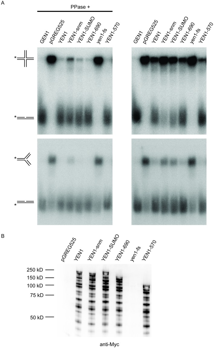
Resolution of branched DNA structures is pivotal for repair of stalled replication forks and meiotic recombination intermediates. The Yen1 nuclease cleaves both Holliday junctions and replication forks. We show that Yen1 interacts physically with Uls1, a suggested SUMO-targeted ubiquitin ligase that also contains a SWI/SNF-family ATPase-domain. Yen1 is SUMO-modified in its noncatalytic carboxyl terminus and DNA damage induces SUMOylation. SUMO-modification of Yen1 strengthens the interaction to Uls1, and mutations in SUMO interaction motifs in Uls1 weakens the interaction. However, Uls1 does not regulate the steady-state level of SUMO-modified Yen1 or chromatin-associated Yen1. In addition, SUMO-modification of Yen1 does not affect the catalytic activity in vitro. Consistent with a shared function for Uls1 and Yen1, mutations in both genes display similar phenotypes. Both uls1 and yen1 display negative genetic interactions with the alternative HJ-cleaving nuclease Mus81, manifested both in hypersensitivity to DNA damaging agents and in meiotic defects. Point mutations in ULS1 (uls1K975R and uls1C1330S, C1333S) predicted to inactivate the ATPase and ubiquitin ligase activities, respectively, are as defective as the null allele, indicating that both functions of Uls1 are essential. A micrococcal nuclease sequencing experiment showed that Uls1 had minimal effects on global nucleosome positioning/occupancy. Moreover, increased gene dosage of YEN1 partially alleviates the mus81 uls1 sensitivity to DNA damage. We suggest a preliminary model in which Uls1 acts in the same pathway as Yen1 to resolve branched DNA structures.


Conflict of interest statement

The authors have declared that no competing interests exist.

Figures

Fig 1
Fig 1. Yen1 SUMOylation is induced by DNA damage and unaffected by uls1Δ and slx5Δ.
(A) Immuno-blot analysis detecting Yen1-13xMyc SUMOylation with an anti-Myc antibody. SUMO conjugated proteins isolated using Ni-NTA agarose from cells with Yen1-13xMyc tagged/untagged at the endogenous locus. The genomic copies of ULP1 and SMT3 were deleted, and the strain contained a plasmid encoding a His6-Flag-tagged SUMO protein. Analysis was performed in WT, uls1Δ and slx5Δ uls1Δ strains in the absence or presence of 0.3% MMS, as indicated. (B) Two-hybrid analysis of the interactions using the indicated bait and prey plasmids. Cells were spotted as 10-fold serial dilutions on indicated SC selection plates and grown for 3 days at 30 °C.
Fig 2
Fig 2. SUMO-modification and the carboxyl terminus are dispensable for Yen1 catalytic activity.
(A) In vitro protein activity assay. SAY1515 (yen1Δ) was transformed with pGREG525 (empty vector) or pGREG525 containing the indicated alleles of YEN1 or human GEN1. 32P-labeled Holliday junction or replication fork DNA substrates were incubated with whole cell extracts containing the indicated myc-tagged alleles for 60 min. The products were analyzed by electrophoresis in a 10% native gel. The substrate and products are schematically shown on the left. (B) Protein blot analysis detecting the expression of the indicated myc-tagged Yen1 proteins used in (A) using an anti-myc antibody.
Fig 3
Fig 3. SIMs in Uls1 are important for the interaction to Yen1.
(A) Schematic diagram of the Uls1 protein. The RING finger domain (red), Helicase domain (grey) and putative SUMO interaction motifs (green), are indicated. The conserved Cys and His residues in the RING finger are shown in red. The Lys residue (K975) in the helicase domain is shown in blue. The residues in the SIMs that were deleted are shown in green. (B) Protein-blot analysis detecting steady state levels of Yen1-TAP, using an anti-TAP antibody. Proteins from whole cell extracts (WCE) and proteins associated with the chromatin fraction was analyzed. An anti-histone H3 antibody was used as loading control. Right panel depicts the average Yen1-Tap/histone H3 ratios from two independent experiments. The ratios in WCE and chromatin fractions are shown defining the ratio in WT as 1. (C) Two-hybrid analysis of the interactions using the indicated bait and prey plasmids. Cells were spotted as 10-fold serial dilutions on indicated SC selection plates and grown for 3 days at 30°C. (D) as in (C), but using truncated Yen1 as bait. (E) Schematic drawing displaying the catalytic N-terminus, the SUMO-modified domain and the Uls1-interaction domain of Yen1.
Fig 4
Fig 4. Genetic interactions among strains lacking Uls1, Mus81 and Yen1.
(A) 10-fold serial dilutions of the wild type parental strain SAY172 and strains containing yen1Δ, uls1Δ, mus81Δ, uls1Δ yen1Δ, mus81Δ yen1Δ, mus81Δ uls1Δ and mus81Δ uls1Δ yen1Δ mutations were spotted on YEPD and YEPD plates with the indicated concentrations of MMS/HU. Cells were grown for 3 days at 30°C. (B) as in (A), but the assay was performed with strains containing uls1Δ, mus81Δ, mus81Δ uls1Δ, mus81Δ uls1K975R-13xMyc, mus81Δ uls1C1330S/C1333S-13xMyc, mus81Δ ULS1-13xMyc, and ULS1-13xMyc mutations. (C) As in (A), but the mus81Δ and mus81Δ uls1Δ strains were transformed with low copy-number plasmids pRS415 and pRS416, pRS416-ULS1 (pJ97) and pRS415-YEN1 (pJ14), as indicated.
Fig 5
Fig 5. Uls1Δ exacerbates the sporulation defect of mus81Δ.
Bar-graph representation of percentage sporulation (A) and spore viability (B) in homozygous diploids with the indicated genotypes. Diploid cells were incubated for 3 days in minimal sporulation media and were analyzed under the microscope for formation of meiotic spores. At least 300 cells were counted for each genotype. Spore viability was assessed by tetrad dissection, analyzing 20 tetrads of each genotype.
Fig 6
Fig 6. Uls1Δ exacerbates the nuclear segregation defect of mms4-mn and is suppressed by spo11Δ.
(A) The indicated strains were transferred to minimal sporulation medium. At the indicated time points, samples were fixed and stained with DAPI. The ratio of cells containing 2 or 4 nuclear masses to total number of cells was determined microscopically in at least 300 cells per genotype. (B)—(D) as in (A), except for the comparison of the genotypes used in (A) to the same strains containing a deletion of SPO11. (E) Uls1Δ had normal levels of phospho-Yen1. Whole cell extracts prepared from SAY172, SAY1506, SAY1558 and W2682 by alkaline lysis were analyzed by SDS-PAGE on a 7.5% acrylamide gel containing 10μM Phos-tag and probed with anti-Myc antibody.
Fig 7
Fig 7. Impact of Uls1 on nucleosomes in the rDNA.
(A) Integrated genome viewer (igv) snapshot displaying the rDNA locus (two repeats). Nucleosome occupancies in WT, sir2 and uls1 strains plotted as blue peaks in the three middle tracks. Nucleosome differential signals between uls1-WT and sir2-WT are the top two tracks. Negative values represent lower nucleosome occupancy in the mutant strains and vice versa. Note that the differential tracks are statistical representations (-log10 (P-value) of Poisson tests, [35]) of the nucleosome differences and that the scales are different. Bottom tracks show the rDNA genes and the amplicons used to determine rDNA copy number. (B) The results of quantitative PCR using four sets of primers amplifying rDNA. Plotted is the average uls1/WT ratio for the E-PRO, ARS, COD and ENH amplicons from two independent experiments, with the variation indicated as error bars. ACT1 was used for normalizing.

References

    1. Clapier CR, Cairns BR. The biology of chromatin remodeling complexes. Annu Rev Biochem. 2009;78:273–304. Epub 2009/04/10. 10.1146/annurev.biochem.77.062706.153223 . - DOI - PubMed
    1. Mohrmann L, Verrijzer CP. Composition and functional specificity of SWI2/SNF2 class chromatin remodeling complexes. Biochim Biophys Acta. 2005;1681(2–3):59–73. Epub 2005/01/04. 10.1016/j.bbaexp.2004.10.005 . - DOI - PubMed
    1. Kingston RE, Narlikar GJ. ATP-dependent remodeling and acetylation as regulators of chromatin fluidity. Genes Dev. 1999;13(18):2339–52. Epub 1999/09/29. . - PubMed
    1. Laurent BC, Treich I, Carlson M. The yeast SNF2/SWI2 protein has DNA-stimulated ATPase activity required for transcriptional activation. Genes Dev. 1993;7(4):583–91. Epub 1993/04/01. . - PubMed
    1. Zhang Z, Buchman AR. Identification of a member of a DNA-dependent ATPase family that causes interference with silencing. Mol Cell Biol. 1997;17(9):5461–72. Epub 1997/09/01. . - PMC - PubMed

Publication types

MeSH terms

LinkOut - more resources