. 2019 Mar 21;15(3):e1007659.
doi: 10.1371/journal.ppat.1007659. eCollection 2019 Mar.

Structural determination of the complement inhibitory domain of Borrelia burgdorferi BBK32 provides insight into classical pathway complement evasion by Lyme disease spirochetes

Affiliations

Structural determination of the complement inhibitory domain of Borrelia burgdorferi BBK32 provides insight into classical pathway complement evasion by Lyme disease spirochetes

Jialei Xie et al. PLoS Pathog. .

Abstract

The carboxy-terminal domain of the BBK32 protein from Borrelia burgdorferi sensu stricto, termed BBK32-C, binds and inhibits the initiating serine protease of the human classical complement pathway, C1r. In this study we investigated the function of BBK32 orthologues of the Lyme-associated Borrelia burgdorferi sensu lato complex, designated BAD16 from B. afzelii strain PGau and BGD19 from B. garinii strain IP90. Our data show that B. afzelii BAD16-C exhibits BBK32-C-like activities in all assays tested, including high-affinity binding to purified C1r protease and C1 complex, and potent inhibition of the classical complement pathway. Recombinant B. garinii BGD19-C also bound C1 and C1r with high-affinity yet exhibited significantly reduced in vitro complement inhibitory activities relative to BBK32-C or BAD16-C. Interestingly, natively produced BGD19 weakly recognized C1r relative to BBK32 and BAD16 and, unlike these proteins, BGD19 did not confer significant protection from serum killing. Site-directed mutagenesis was performed to convert BBK32-C to resemble BGD19-C at three residue positions that are identical between BBK32 and BAD16 but different in BGD19. The resulting chimeric protein was designated BXK32-C and this BBK32-C variant mimicked the properties observed for BGD19-C. To query the disparate complement inhibitory activities of BBK32 orthologues, the crystal structure of BBK32-C was solved to 1.7Å limiting resolution. BBK32-C adopts an anti-parallel four-helix bundle fold with a fifth alpha-helix protruding from the helical core. The structure revealed that the three residues targeted in the BXK32-C chimera are surface-exposed, further supporting their potential relevance in C1r binding and inhibition. Additional binding assays showed that BBK32-C only recognized C1r fragments containing the serine protease domain. The structure-function studies reported here improve our understanding of how BBK32 recognizes and inhibits C1r and provide new insight into complement evasion mechanisms of Lyme-associated spirochetes of the B. burgdorferi sensu lato complex.


Conflict of interest statement

The authors have declared that no competing interests exist.

Figures

Fig 1
Fig 1. BBK32 orthologues are encoded by Borrelia burgdorferi sensu lato isolates.
A) BBK32 is a multifunctional lipoprotein expressed on the surface of B. burgdorferi. BBK32 interacts with three vertebrate host macromolecules via non-overlapping binding sites. The intrinsically disordered N-terminal domain of BBK32 (BBK32-N) recognizes certain glycosaminoglycans and the human extracellular matrix protein fibronectin, while the globular C-terminal region (BBK32-C) binds to the complement protease C1r within the C1 complex. B) A sequence alignment of the C-terminal domain of BBK32 orthologues from the Lyme disease-associated spirochetes B. burgdorferi BBK32 from strain B31, B. garinii BGD19 from strain IP90, and B. afzelii BAD16 from strain PGau is shown. Residues selected for mutational analysis in this study (i.e. “BXK32-C”) are highlighted in yellow and marked with arrows.
Fig 2
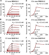
Fig 2. The C-terminal domain of BGD19 and BAD16 bind with high affinity to human C1 and C1r.
The ability of the C-terminal region of BGD19 (BGD19-C) and BAD16 (BAD16-C) to bind human C1 or C1r, was assessed by SPR. BBK32-C was used as a control. For C1, a two-fold dilution series (0.1 to 150 nM) was injected over immobilized BBK32-C (panel A), BGD19-C (panel B), and BAD16-C (panel C). The raw sensorgrams are drawn as black lines and the results of kinetic fitting analysis using Biacore T200 Evaluation Software are drawn as red lines. For C1r the a single-cycle analysis was performed using a five-fold dilution series (1.6 to 1000 nM) of BBK32-C (panel D), BGD19-C (panel E), and BAD16-C (panel F). For clarity, the dissociation phase of each sensorgram is labeled with the C1r injection concentration. Sensorgrams from a representative injection series are shown and all experiments were conducted in triplicate with the dissociation constants (KD) reported as the mean ± S.D.
Fig 3
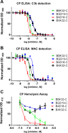
Fig 3. BGD19 and BAD16 inhibit the classical pathway of complement.
A) Two in vitro assays of classical pathway complement activation were used to assess the relative inhibitory activity of recombinant BBK32-C, BGD19-C, BAD16-C, and BXK32-C. (A-B) an ELISA-based assay was used in the presence of a two-fold concentration series of BBK32-C, BGD19-C, BAD16-C, and BXK32-C (1 nM to 2,000 nM). A) C3b deposition or B) MAC deposition was detected in separate experiments each performed in duplicate. C) A classical pathway-specific hemolytic assay was used in the presence of a concentration series of each inhibitor (31 to 1,000 nM) to assess the relative ability of each protein to protect sensitized sheep red blood cells from complement-mediated lysis in 1% normal human serum. Each experiment was performed in triplicate and values are reported as the mean ± SEM.
Fig 4
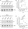
Fig 4. Binding of C1 and C1r to BBK32 orthologues via far western blot analysis.
A-D) BGD19, BBK32, and BAD16 were expressed as lipoproteins on the surface of B. burgdorferi B314. Whole cell protein lysates were separated on an SDS-PAGE gel and probed for binding to human C1 (panel A) or C1r (panel C) using a Far Western blot overlay. Samples tested include strain B314/pBBE22luc (vector only control; labeled as “Vector”), B314/pBDG19 (labeled as BGD19), B314/pCD100 (labeled as BBK32), B314/pBAD16 (labeled as BAD16), and B314 alone (labeled as null). FlaB was used as a loading control to normalize variation between C1 and C1r binding by BBK32, BAD16, and BGD19 in panels A and C. Densitometry was performed from independent blots to quantify the observed signals as depicted in panels A and C. Panels B and D report the signal detected for C1 and C1r to the samples indicated on the x axis, respectively. All values were normalized relative to BBK32 binding to either C1 or C1r. P values between samples are indicated above the bars.
Fig 5
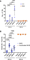
Fig 5. Native BAD16 and BGD19 exhibit differential binding to C1 and C1r and confer serum-resistance when expressed on the surface of spirochetes.
A) Natively expressed bad16 (labeled as BAD16) and bgd19 (labeled as BGD19), were tested for their ability to bind immobilized C1r (blue circles) or BSA (yellow squares) relative to B314 containing BBK32 (labeled as BBK32) or B314 with vector DNA alone (labeled as Vector). Binding was done in triplicate for independent samples and the average and standard deviation shown. B) The ability of each protein to confer resistance to normal human serum (NHS) was assessed in the serum sensitive B. burgdorferi strain B314. Sensitivity was scored as a ratio of the affected cells relative to the total cells viewed. Cells affected were categorized as those that lacked motility, exhibited membrane damage, or manifested overt cell lysis (blue circles). Heat inactivated NHS was used as a control and is shown on the right (yellow squares). P values between samples are indicated above the bars. ns, not significant.
Fig 6
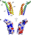
Fig 6. The crystal structure of the complement inhibitory domain of BBK32.
A) The structure of BBK32(206–348) solved at 1.7Å resolution (PDB: 6N1L). A ribbon diagram representation using a spectrum-based coloration scheme of BBK32(206–348) where the N-terminal region of the protein is colored in blue and the C-terminus in red. The structure is shown turned 180° about the y-axis. BBK32(206–348) is characterized by a helical bundle fold where helices 1, 3, 4, and 5 form a core four-helix bundle motif and helix 2 extends away from the core at ~120° relative to helix 3. B) BBK32 is drawn in a surface representation in the same orientations as depicted in panel A. The Adaptive Poisson-Boltzmann Solver as implemented in Pymol was used to calculate the electrostatic potential of the molecular surface. The color scheme represents a gradient of electrostatic potential where regions of negative (red) and positive (blue) are contoured at ± 2 kbT/e where kb is Boltzmann’s constant = 1.3806 x 10⁻²³ J K-1, T is temperature in K, and e is the charge of an electron = 1.6022 x10⁻¹⁹ C.
Fig 7
Fig 7. Residues in the BBK32 to BGD19 chimera are solvent exposed.
A) A chimeric BBK32-C protein encoding three charged residues which are identical between BBK32 and BAD16, but different in BGD19-C, exhibits BDG19-like activity (see sequence alignment in Fig 1B and Fig 3). The structure of BBK32-C is oriented to highlight each of these residues (colored orange, stick representation). B) A molecular surface representation of BBK32-C in the same orientation as shown in panel A indicates all three residues altered in the BXK32-C chimera construct are surface exposed in the BBK32-C crystal structure.
Fig 8
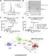
Fig 8. BBK32 binds to the C-terminal region of C1r and requires the SP domain for high affinity interaction.
A-B) Intrinsic proteolysis of C1r reaches completion upon overnight incubation at 37°C resulting in the release of a fragment corresponding to the C-terminal domains CCP1-CCP2-SP. The auto-catalyzed digestion reaction of C1r was injected onto a size exclusion column. The C1rCCP1-CCP2-SP-auto proteolytic fragment elutes in peak 2. When BBK32(206–348) is added to the C1r digestion reaction at 2-fold molar excess (relative to full-length C1r) a new peak appeared, peak 3, which contains both BBK32(206–348) and the C1rCCP1-CCP2-SP-auto proteolytic fragment, as judged by mass spectrometry analysis. C) To confirm that BBK32 recognizes the C-terminal C1r CCP-1-CCP2-SP domains, SPR binding studies were performed. Purified C1rCCP1-CCP2-SP-auto exhibited high affinity interaction for BBK32(206–348) (KD = 1.5 nM) D) Recombinant refolded His-C1r-CCP2-SP retains high affinity interaction (KD = 3.9 nM), whereas recombinant His-CCP1 or His-CCP1-CCP2 alone fail to interact with BBK32(206–348). E) A model of full-length C1r is shown which is built from the available crystal structures of C1r domain truncations (PDB’s: 4LOT, 6F39, and 1GPZ). The location of the C1rCCP1-CCP2-SP-auto proteolytic fragment is indicated. Together these data indicate that BBK32 targets the C-terminal region of the C1r protease and requires the SP domain for high-affinity interaction.

Similar articles

Cited by

References

    1. Kuehn BM. CDC Estimates 300 000 US Cases of Lyme Disease Annually. JAMA. 2013;310: 1110 10.1001/jama.2013.278331 - DOI - PubMed
    1. Margos G, Vollmer SA, Ogden NH, Fish D. Population genetics, taxonomy, phylogeny and evolution of Borrelia burgdorferi sensu lato. Infect Genet Evol. 2011;11: 1545–1563. 10.1016/j.meegid.2011.07.022 - DOI - PMC - PubMed
    1. Merle NS, Church SE, Fremeaux-Bacchi V, Roumenina LT. Complement System Part I—Molecular Mechanisms of Activation and Regulation. Front Immunol. 2015;6: 262 10.3389/fimmu.2015.00262 - DOI - PMC - PubMed
    1. Merle NS, Noe R, Halbwachs-Mecarelli L, Fremeaux-Bacchi V, Roumenina LT. Complement System Part II: Role in Immunity. Front Immunol. 2015;6: 257 10.3389/fimmu.2015.00257 - DOI - PMC - PubMed
    1. Lambris JD, Ricklin D, Geisbrecht BV. Complement evasion by human pathogens. Nature Reviews Microbiology. 2008;6: 132–142. 10.1038/nrmicro1824 - DOI - PMC - PubMed

Publication types

MeSH terms