. 2019 Mar 21;14(3):e0214275.
doi: 10.1371/journal.pone.0214275. eCollection 2019.

Genetic and epigenetic profiling of the infertile male

Affiliations

Genetic and epigenetic profiling of the infertile male

Stephanie Cheung et al. PLoS One. .

Abstract

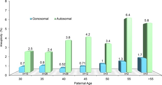
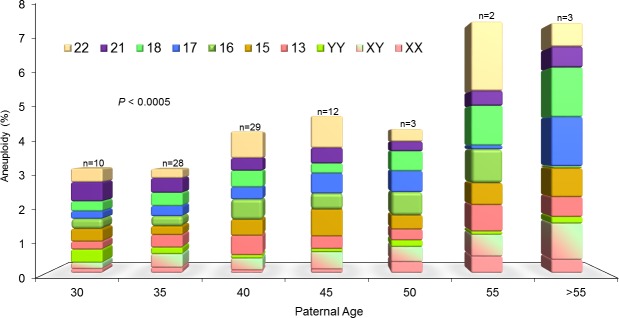
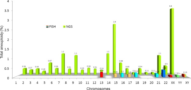
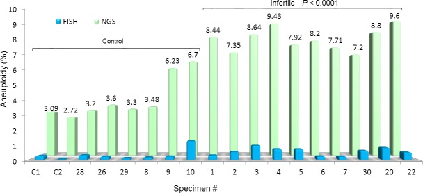
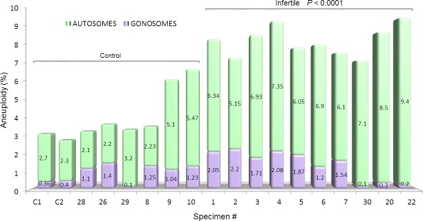
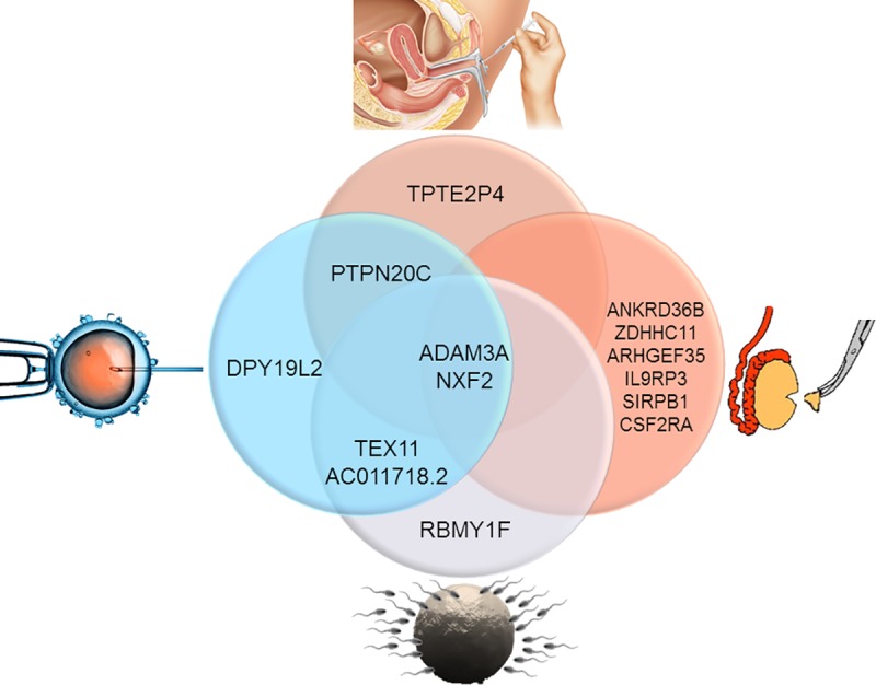
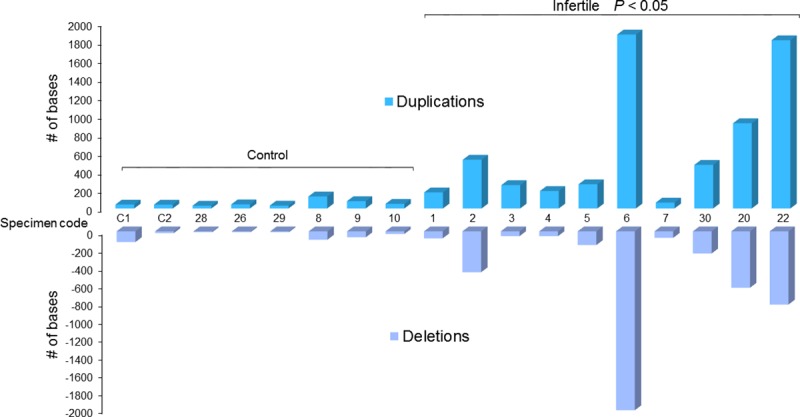
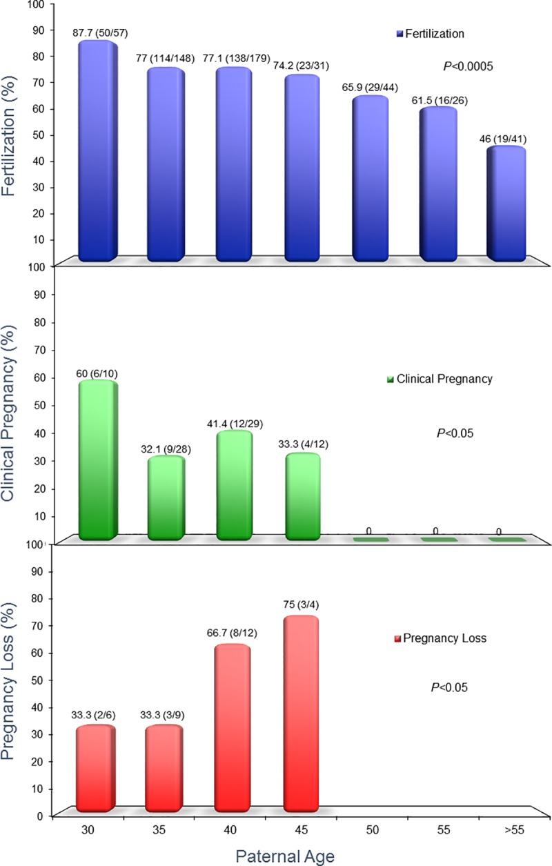
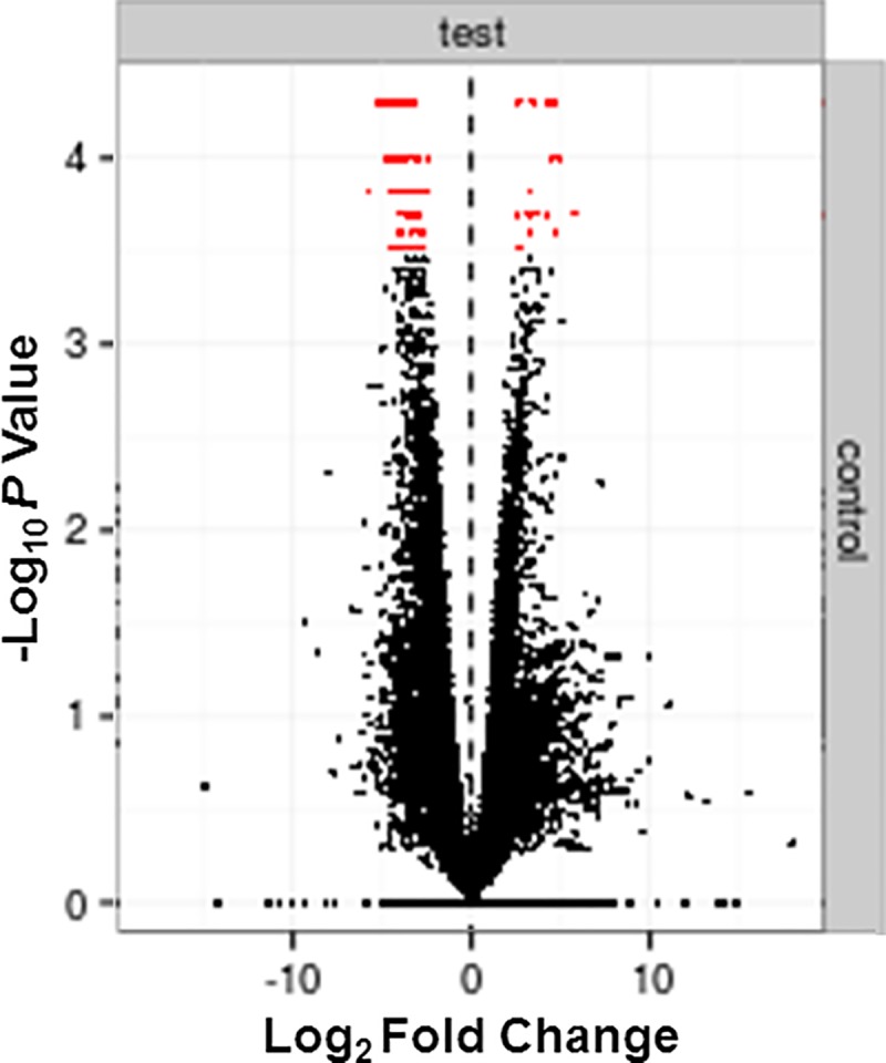
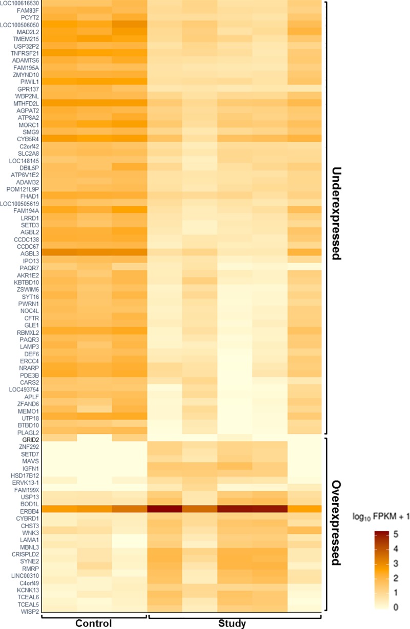
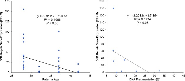
Evaluation of reproductive quality of spermatozoa by standard semen analysis is often inadequate to predict ART outcome. Men may be prone to meiotic error and have higher proportion of spermatozoa with fragmented chromatin, capable of affecting the conceptus' health. In men with unexplained infertility, supplementary tests may be pivotal to gain insight into the paternal contribution to the zygotic genome. A total of 113 consenting men were included in the study, with an additional 5 donor specimens used as control. Among study participants, 87 were screened for sperm aneuploidy by fluorescent in situ hybridization (FISH) and ranked according to their increasing age. A total of 18 men were assessed by whole exome sequencing and categorized according to their reproductive outcome as either fertile or infertile. Another set of men (n = 13) had their gene expression analyzed by RNA-seq and were profiled according to their reproductive capacity. FISH revealed that the average aneuploidy rate was highest for men over-55 age group (9.6%), while men >55 had the highest average disomy for chromosomes 17(1.2%) and 18(1.3%). ART results for the entire cohort comprised 157 cycles, stratified by paternal age. The youngest age group (25-30 years) had a fertilization rate of 87.7% which decreased to 46.0% in the >55 age group. Clinical pregnancy rate was highest in the 25-30yr group (80.0%) while no pregnancies were attained in the >55 age groups. Pregnancy loss was characterized by a steadily increasing trend, highest in the 51-55 age group (50.0%). NGS was performed on a cohort of patients classified as having recurrent pregnancy loss. This cohort was classified as the infertile group (n = 10) and was compared to a control group (n = 8) consisting of patients successfully treated by ART. Eight couples in 17 ICSI cycles achieved a clinical pregnancy rate of 82.4% while 10 infertile couples treated in 21 cycles achieved a pregnancy rate of 23.8%, all resulting in pregnancy loss. DNA-sequencing on spermatozoa from these patients yielded overall aneuploidy of 4.0% for fertile and 8.6% for the infertile group (P<0.00001). In the infertile cohort, we identified 17 genes with the highest mutation rate, engaged in key roles of gametogenesis, fertilization and embryo development. RNA-seq was performed on patients (n = 13) with normal semen analyses. Five men unable to attain a pregnancy after ART were categorized as the infertile group, while 8 men who successfully sustained a pregnancy were established as the fertile control. Analysis resulted in 86 differentially expressed genes (P<0.001). Of them, 24 genes were overexpressed and 62 were under-expressed in the infertile cohort. DNA repair genes (APLF, CYB5R4, ERCC4 and TNRFSF21) and apoptosis-modulating genes (MORC1, PIWIL1 and ZFAND6) were remarkably under-expressed (P<0.001). Sperm aneuploidy assessment supported by information on gene mutations may indicate subtle dysfunctions of the spermatozoon. Furthermore, by querying noncoding RNA we may gather knowledge on embryo developmental competence of spermatozoa, providing crucial information on the etiology of unexplained infertility of the infertile male.


Conflict of interest statement

The authors have declared that no competing interests exist.

Figures

Fig 1
Fig 1. Sperm aneuploidy assessment by FISH depicting sperm chromosomal defects.
A comparison between autosomal aneuploidy and male age, assesed by Pearson's Correlation Coefficient, had an R2 value of 0.6 (P<0.00001). Data has been allocated into different age bins, as shown, for clinical relevance and visual representation.
Fig 2
Fig 2. FISH aneuploidy assessment by individual chromosomes in relation to advancing male age.
Of particular note were chromosomes 15 (orange) and 17 (blue), which progressively increased with age, while chromosome 21 (purple) was noticeably highest in the youngest age group, progressively decreasing with advancing male age.
Fig 3
Fig 3. Assessment of sperm aneuploidy for each chromosome by two different methods.
In the first row, the minute multicolor columns indicate the chromosomal defects assessed by FISH on 9 chromosomes. In contrast, the green histogram represents whole genome molecular karyotyping by NGS. This technique allows for a more accurate and comprehensive assessment of all chromosomes to detect sperm aneuploidy.
Fig 4
Fig 4. Aneuploidy assessment by FISH and NGS in the study population.
FISH analysis, depicted by the blue histogram, failed to evidence any difference among the groups. NGS, however, reported a remarkable and significant aneuploidy rate in the infertile group when compared to the fertile control (P<0.0001).
Fig 5
Fig 5. NGS assessment of sperm aneuploidy according to chromosome type in the study population.
Both autosomal and gonosomal chromosomes contributed to the overall higher aneuploidy of the infertile cohort in comparison to the fertile control group (P<0.0001). The most represented chromosomal defects were related to the autosomes (green portion of the histogram).
Fig 6
Fig 6. Gene mutation assessment according to ART treatment.
Several gene mutation commonalities were identified when taking into consideration the type of assisted reproduction technique used. In couples treated by IUI, only a pseudogene (TPTE2P4) was identifed while a gene involved in spermatogenesis (RBMY1F) were mutated in those treated by IVF. A gene supporting sperm acrosome formation (DPY19L2) was mutated in men requiring ICSI. When we assessed gene mutations in men treated by testicular biopsy, a variety of genes were identified that play a role in basic cellular functions (ANKRD36B), androgen production modulation (SIRPB1), or activation of germ cells (CSF2RA). Common gene mutations were also identified in patients who underwent multiple types of ART treatments. For instance, men treated by both IVF and ICSI shared an imbalance of TEX11, a gene involved in meiotic crossing-over. All couples possessed gene mutations in ADAM3A and NXF2, which support sperm-egg fusion and RNA nucleus-cytoplasmic transport, respectively.
Fig 7
Fig 7. Histogram graph displaying overall gene mutations observed by CNV analysis in the study population.
Each bar indicates a specimen. The gene duplications (dark blue histogram) and gene deletions (light blue histogram) are higher in the infertile cohort in comparison to the fertile control group (P<0.05).
Fig 8
Fig 8. Fertilization, clinical pregnancy, and rate of pregnancy loss in relation to paternal age.
ICSI outcome was assessed for men whom we performed a FISH assessment on. Fertilization was characterized by a decreasing trend and was lowest in the >55 age group. Clinical pregnancy rates was highest in the 25–30 age group but became absent in the 51–55 and >55 age groups. The rate of pregnancy loss was also characterized by an increasing trend with advancing paternal age.
Fig 9
Fig 9. Volcano plot indicating the significantly imbalanced (in red) genes in logarithmic fold of RPKM.
We identified a total of 24 over-expressed genes on the right of the dotted line, and 62 under-expressed genes on the left.
Fig 10
Fig 10. Heat map depicting the differentially expressed genes (by RNA analysis in spermatozoa) in the study group versus the reference control.
Within the study group, there were 24 over-expressed (bottom right) and 62 under-expressed genes (top right) identified.
Fig 11
Fig 11. Effect of paternal age and DNA fragmentation on DNA repair gene expression.
Assessment of the 7 most significant differentially expressed genes according to paternal age (left graph) showed an inverse correlation, with the oldest men having much lower expression of these DNA repair and apoptotic regulating genes (P<0.05). Similarly, plotting the expression of these genes in function of increasing sperm DNA fragmentation (right graph) illustrated that the higher the expression of genes related to DNA repair and apoptosis, the lower the sperm DNA fragmentation (P<0.05).

Similar articles

Cited by

References

    1. Boivin J, Bunting L, Collins J, Nygren K. International estimates of infertility prevalence and treatment-seeking: potential need and demand for infertility medical care. Human Reproduction. 2007;22(6):6 Epub 2007, March 21. 10.1093/humrep/dem046. - DOI - PubMed
    1. Sabanegh ES. Male Infertility: Probloems and Solutions Eric A. Klein MD, editor. Cleveland, Ohio: Springer Science+Business Media, LLC; 2011.
    1. World Health Organization. WHO laboratory manual for the Examination and processing of human semen. 5th ed. Switzerland2010.
    1. AUA TMIBPPCotAUAPCotASfRM. Report on optimal evaluation of the infertile male. Fertility and Sterility. 2006;82(5). 10.1016/j.fertnstert.2006.08.029. - DOI - PubMed
    1. Van Den Hoven L, Hendriks JC, Verbeet JG, Westphal JR, Wetzels AM. Status of sperm morphology assessment: an evaluation of methodology and clinical value. Fertil Steril. 2015;103(1):53–8. Epub 2014/12/03. 10.1016/j.fertnstert.2014.09.036 . - DOI - PubMed

MeSH terms