Review
. 2019 May:77:76-86.
doi: 10.1016/j.dnarep.2019.03.004. Epub 2019 Mar 7.

Mechanisms underlying aflatoxin-associated mutagenesis - Implications in carcinogenesis

Affiliations
Review

Mechanisms underlying aflatoxin-associated mutagenesis - Implications in carcinogenesis

Amanda K McCullough et al. DNA Repair (Amst). 2019 May.

Abstract

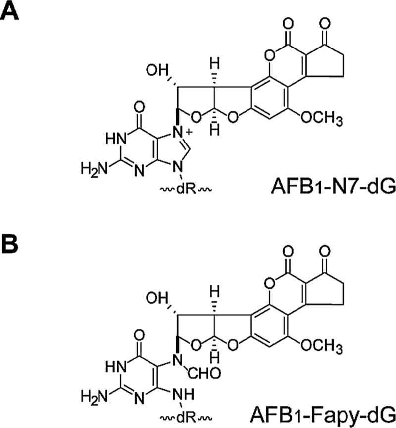
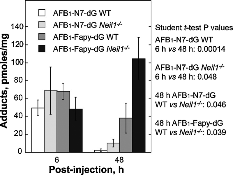
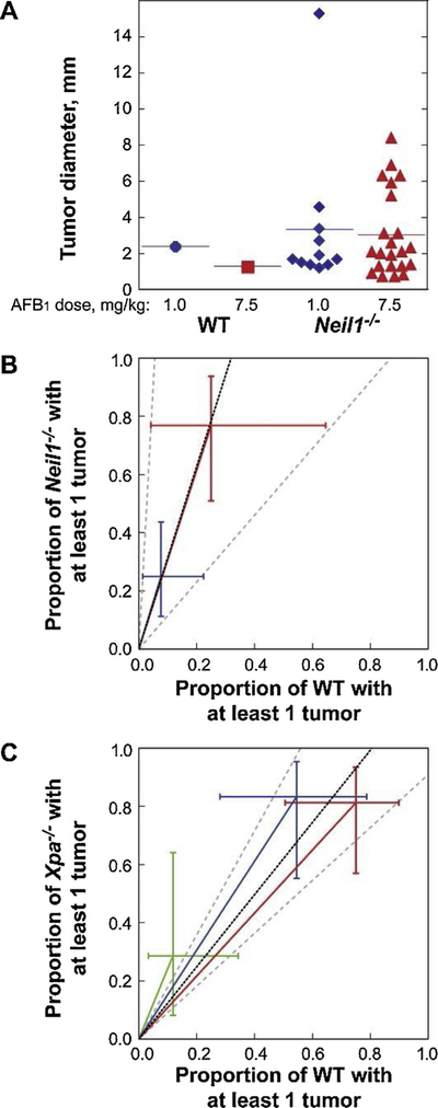
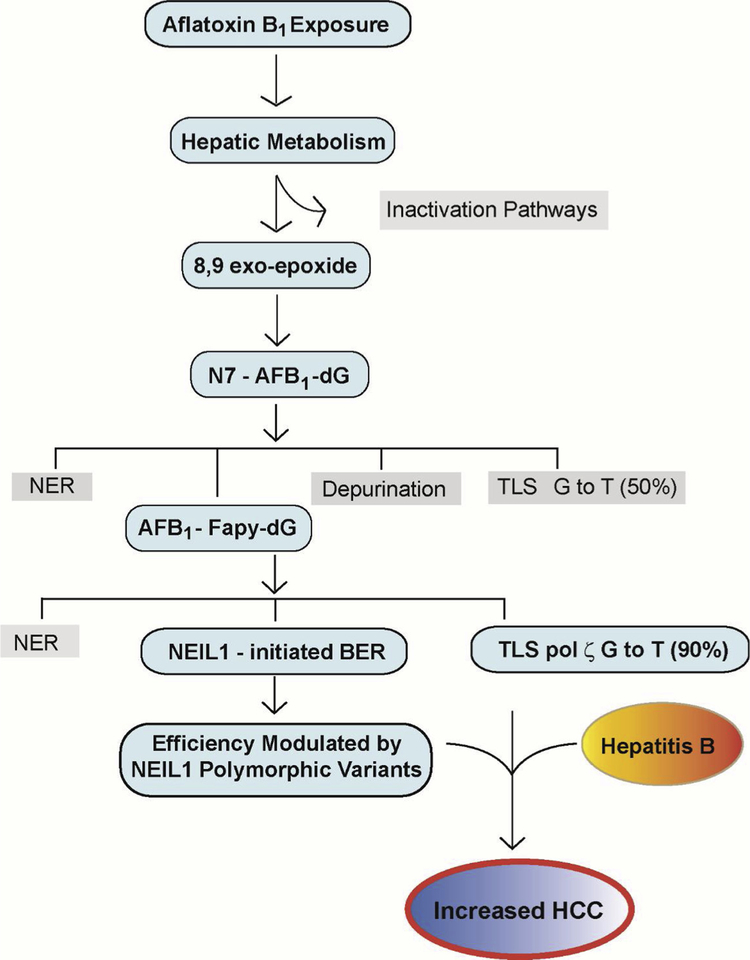
Chronic dietary exposure to aflatoxin B1 (AFB1), concomitant with hepatitis B infection is associated with a significant increased risk for hepatocellular carcinomas (HCCs) in people living in Southeast Asia and sub-Saharan Africa. Human exposures to AFB1 occur through the consumption of foods that are contaminated with pervasive molds, including Aspergillus flavus. Even though dietary exposures to aflatoxins constitute the second largest global environmental risk factor for cancer development, there are still significant questions concerning the molecular mechanisms driving carcinogenesis and what factors may modulate an individual's risk for HCC. The objective of this review is to summarize key discoveries that established the association of chronic inflammation (most commonly associated with hepatitis B viral (HBV) infection) and environmental exposures to aflatoxin with increased HCC risk. Special emphasis will be given to recent investigations that have: 1) refined the aflatoxin-associated mutagenic signature, 2) expanded the DNA repair mechanisms that limit mutagenesis via adduct removal prior to replication-induced mutagenesis, 3) implicated a specific DNA polymerase in the error-prone bypass and resulting mutagenesis, and 4) identified human polymorphic variants that may modulate individual susceptibility to aflatoxin-induced cancers. Collectively, these investigations revealed that specific sequence contexts are differentially resistant against, or prone to, aflatoxin-induced mutagenesis and that these associations are remarkably similar between in vitro and in vivo analyses. These recent investigations also established DNA polymerase ζ as the major polymerase that confers the G to T transversion signature. Additionally, although the nucleotide excision repair (NER) pathway has been previously shown to repair aflatoxin-induced DNA adducts, recent murine data demonstrated that NEIL1-initiated base excision repair was significantly more important than NER relative to the removal of the highly mutagenic AFB1-Fapy-dG adducts. These data suggest that inactivating polymorphic variants of NEIL1 could be a potential driver of HCCs in aflatoxin-exposed populations.

Keywords: DNA repair; DNA replication; Hepatocellular carcinoma; NEIL1; Polymerase zeta.


Conflict of interest statement

Conflict of interest

The authors declare that there are no conflicts of interest.

Figures

Fig. 1.
Fig. 1.. DNA Adduct Structures.
(A) Structure of the AFB1-N7-dG adduct. (B) Structure of the AFB1-Fapy-dG adduct. [Reproduced from [76] Fig. 1].
Fig. 2.
Fig. 2.. Mammalian pol ζ protects against AFB1-induced cytotoxicity.
(A) Growth inhibition in Rev3L+/− (circles), Rev3L−/− (squares), and Rev3L−/− +hRev3L (triangles) MEFs was determined 48 h after AFB1 treatment by measuring cellular ATP. (B) Quantification of total cell death (Annexin V+/PI-, Annexin V-/PI + and Annexin V+/PI+) in Rev3L+/− (black bars) and Rev3L−/− (gray bars) MEFs following AFB1 treatment. Data represent mean ± SEM from three independent experiments. * P < 0.05 by unpaired two-tailed t-test with unequal variances. [Reproduced from [57], Fig. 1, Panels A and B].
Fig. 3.
Fig. 3.. Genome instability generated by AFB1 exposures in pol ζ-deficient cells.
(A) DSB formation was assessed by measuring the accumulation and resolution of γ-H2AX foci in response to 200 nM AFB1 +S9 for 1.5 h. Representative images of γ-H2AX foci in Rev3L+/− (top) and Rev3L−/− (bottom) cells at 72 h post-exposure. (B) Quantification of γ-H2AX foci over time in response to AFB1 exposure. (C–F) Genome instability in interphase cells was assessed by micronuclei formation and the appearance of multinucleated cells. Representative images (C) and quantification (D) of micronuclei. Representative images (E) and quantification (F) of multinucleated cells. (G–H) Following a 1.5 h AFB1 exposure, genome instability in metaphase cells was assessed by chromosome breakage analyses after 48 h recovery. Quantification of chromatid breaks (G) and chromosomal radials (H) reflecting aberrant DNA repair. Data represent the mean ± SEM from three independent experiments. * p < 0.05, ** p < 0.01, *** p < 0.001 by unpaired two-tailed t-test with unequal variances. The p-values above bars are comparing treated and untreated cells; p-values between bars are comparing between cell lines. n/s = not significant. Black bars = Rev3L+/−; Gray bars = Rev3L−/−. [Reproduced from [57], Fig. 3].
Fig. 4.
Fig. 4.. NEIL1-catalyzed incision of DNA containing an AFB1-Fapy-dG adduct.
32P-labeled oligodeoxynucleotides (20 nM) containing either AFB1-Fapy-dG in fully duplex DNA (A) or Tg in fully duplex DNA (B) were incubated with hNEIL1 (230 nM). The aliquots were removed at the indicated times, and following separation by gel-electrophoresis, DNA was visualized using a phosphor screen and Personal Molecular Imager™ System (Bio-Rad). The representative gel images are shown. [Reproduced from [76], Fig. 2].
Fig. 5.
Fig. 5.. Levels of AFB1-induced DNA adducts in liver DNA from of WT and Neil1−/− mice following AFB1 IP injection.
Neil1−/− and control WT mice (6 day old pups) were injected with 10 mM AFB1 in DMSO at a dose of 3.5 mg/kg. Livers were harvested at 6 and 48 h post-injection, immediately frozen in liquid nitrogen, and DNA isolated for AFB1 adduct analysis. Following acid hydrolysis, internal 15N5-guanine-derived standards were added to permit quantitative analysis by isotope dilution mass spectrometry for both AFB1-N7-dG and AFB1-Fapy-dG. Adducts were separated by ultra-performance liquid-chromatography mass spectrometry. The protonated parent ion of the AFB1-N7-dG adduct (m/z 480.1) was selected and subjected to collision-induced fragmentation producing a m/z 152 product ion that was monitored to quantify adduct levels. The AFB1-Fapy-dG adduct was measured by selection of the m/z 498 parent ion and monitoring the collision-induced product ion m/z 452.29. [Reproduced from [76], Fig. 2].
Fig. 6.
Fig. 6.. AFB1-induced carcinogenesis in Neil1−/− and XPA−/− mice.
(A) The individual diameter of liver tumors observed in AFB1-injected WT and Neil1−/− mice. Relative AFB1-induced tumor risk analysis in (B) Neil1−/− mice, with data illustrated by blue and red symbols representing AFB1 doses of 1.0 and 7.5 mg/kg respectively and (C) XPA−/− mice, with data illustrated by green, blue and red symbols representing AFB1 doses of 0, 0.6 and 1.5 mg/kg, respectively. [Reproduced from [76], Fig. 4].
Fig. 7.
Fig. 7.. Key Steps in Modulating Aflatoxin B1 Mutagenesis and Carcinogenesis.
Following ingestion of foods that are contaminated with Aspergillus flavus, hepatic metabolism shuttles AFB1 into pathways that are either biologically harmless (Inactivation Pathways) or biologically activated as the 8,9 exo-epoxide for subsequent reaction at N7-guanine. Although the product of this reaction, the N7- AFB1-dG adduct is relatively short lived, it can be repaired by NER, undergo depurination with repair completed via BER, replicated by an error-prone mechanism yielding ~50% G to T transversions, or hydrolyzed to the long-lived AFB1-Fapy-dG adduct. This adduct is subject to repair via NER, replicated in a highly error-prone mechanism by DNA polymerase ζ, yielding ~90% G to T transversions and ~7% G to A transitions, or repaired via BER initiated by NEIL1. Catalytically-inactive, but accurately folded, polymorphic variants of NEIL1 are hypothesized to interfere with normal repair and thus, lead to increased numbers of unrepaired sites that contribute to overall genetic instability. It is this combination of decreased repair, error-prone replication, and hepatitis B infection and chronic inflammation that leads to increased risk of HCC formation.

Similar articles

Cited by

References

    1. Kensler TW, Roebuck BD, Wogan GN, Groopman JD, Aflatoxin: a 50-year odyssey of mechanistic and translational toxicology, Toxicol. Sci 120 (Suppl 1) (2011) S28–S48. - PMC - PubMed
    1. Kew MC, Aflatoxins as a cause of hepatocellular carcinoma, J. Gastrointestin. Liver Dis 22 (2013) 305–310. - PubMed
    1. Sargeant K, Sheridan A, O’Kelly J, Carnaghan RBA, Toxicity associated with certain samples of groundnuts, Nature 192 (1961) 1096–1097.
    1. Lancaster MC, Jenkins FP, Philip JM, Toxicity associated with certain samples of groundnuts, Nature 192 (1961) 1095–1096.
    1. Blount WP, Turkey “x” disease, Turkeys (J. Br. Turkey Fed.) 9 (1961) 52–58.

Publication types

MeSH terms

LinkOut - more resources