. 2019 Jun:24:101166.
doi: 10.1016/j.redox.2019.101166. Epub 2019 Mar 12.

The vitamin E derivative garcinoic acid from Garcinia kola nut seeds attenuates the inflammatory response

Affiliations

The vitamin E derivative garcinoic acid from Garcinia kola nut seeds attenuates the inflammatory response

Maria Wallert et al. Redox Biol. 2019 Jun.

Abstract

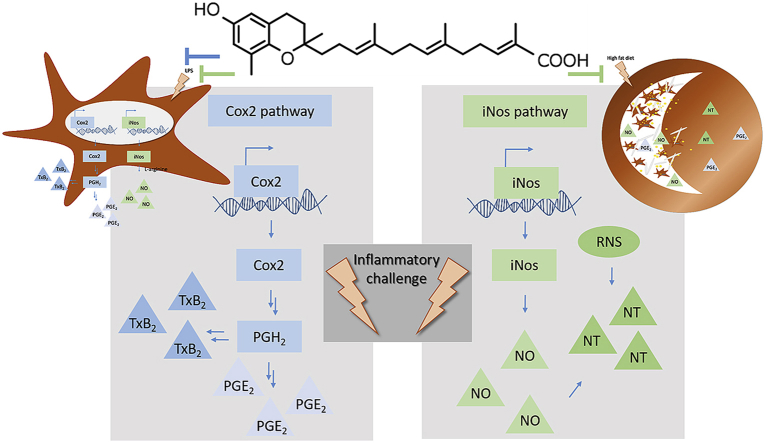
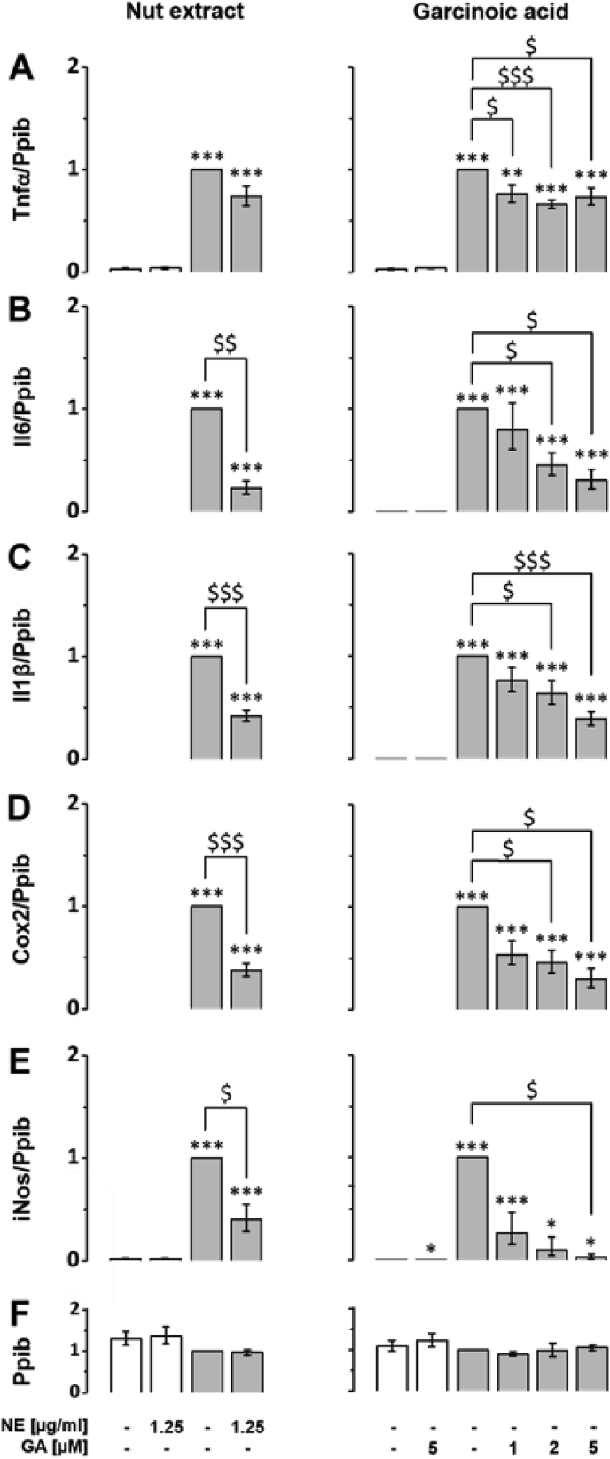
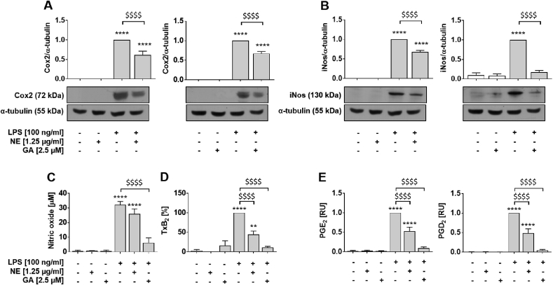
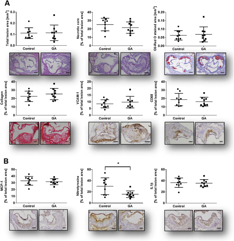
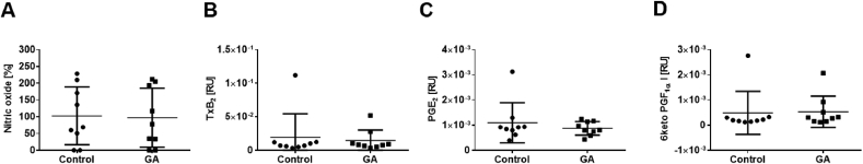
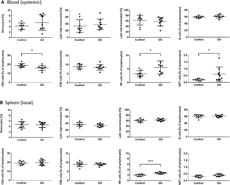
The plant Garcinia kola is used in African ethno-medicine to treat various oxidation- and inflammation-related diseases but its bioactive compounds are not well characterized. Garcinoic acid (GA) is one of the few phytochemicals that have been isolated from Garcinia kola. We investigated the anti-inflammatory potential of the methanol extract of Garcinia kola seeds (NE) and purified GA, as a major phytochemical in these seeds, in lipopolysaccharide (LPS)-activated mouse RAW264.7 macrophages and its anti-atherosclerotic potential in high fat diet fed ApoE-/- mice. This study outlines an optimized procedure for the extraction and purification of GA from Garcinia kola seeds with an increased yield and a purity of >99%. We found that LPS-induced upregulation of iNos and Cox2 expression, and the formation of the respective signaling molecules nitric oxide and prostanoids, were significantly diminished by both the NE and GA. In addition, GA treatment in mice decreased intra-plaque inflammation by attenuating nitrotyrosinylation. Further, modulation of lymphocyte sub-populations in blood and spleen have been detected, showing immune regulative properties of GA. Our study provides molecular insights into the anti-inflammatory activities of Garcinia kola and reveals GA as promising natural lead for the development of multi-target drugs to treat inflammation-driven diseases.

Keywords: Atherosclerosis; Garcinia kola seeds; Garcinoic acid; Inflammatory response; Macrophage activation.


Figures

Image 1
Graphical abstract
Fig. 1
Fig. 1
Bligh and Dyer extraction increased the yield of garcinoic acid (GA) isolated from Garcinia kola seeds at high purity. Representative LC-MS chromatograms of the Garcinia kola seeds extracts obtained by the standard procedure (A) and by Bligh and Dyer extraction (B). Panel (C) and (D) show LC-MS chromatograms of the purified GA obtained from crude methanol extract from Garcinia kola seeds according to the procedures used for (A) and (B), respectively. Mass spectra of the purified GA were obtained from the LC-MS chromatogram (D) for two peaks with retention times of 9.8–10.6 min (main peak, E) and 10.7–11.4 min (minor peak, G). MS/MS fragmentation spectra of (E) and (G) are shown in panels (F) and (H). The fragmentation is indicated on structure (I), respectively.
Fig. 2
Fig. 2
Lipopolysaccharide-induced upregulation of Il6, Il1β, Cox2, iNos and Tnfα mRNA expression is blocked by the NE and isolated GA. RAW264.7 were pre-incubated with either NE (left column) GA (right column) or solvent (DMSO) for 24 h (white bars). For LPS-induced experiments, macrophages were co-incubated with 100 ng/ml LPS and either solvent, NE or GA at the doses indicated for another 24 h (grey bars). Samples co-cultured with solvent and LPS were defined as one. Expression levels of the inflammatory response genes (A) Tnfα, (B) Il6, (C) Il1β, (D) Cox2 and (E) iNos were measured using RT-qPCR and normalized to the mRNA expression of the reference gene (F) peptidylprolyl isomerase B (Ppib). Error bars display calculated minimum and maximum of SEM (SEM ± min, max) expression levels of four independent biological experiments, each measured in one or two technical replicates. *, p < 0.05; **p < 0.01; ***, p < 0.001 (vs. solvent control); $, p < 0.05; $$, p < 0.01; $$$, p < 0.001 (vs. LPS treatment). Student's t-test was performed for statistical analysis.
Fig. 3
Fig. 3
Lipopolysaccharide-induced expression of iNos and Cox2 protein and secretion of respective signaling molecules are more potently blocked by GA compared to NE. RAW264.7 macrophages were incubated with either solvent (DMSO, white bars), NE or GA, or co-incubated with 100 ng/ml LPS (grey bars). The NE and GA decreased the protein expression of (A) Cox2 and (B) iNos after 24 h pre-incubation with NE or GA followed by 14 h and 24 h co-incubation with LPS, respectively. Samples incubated with LPS were defined as reference and were set as one. Protein levels were normalized to α-tubulin for quantification and representative Western blots are shown (for un-chopped versions see Suppl. Fig. S6). (C) Basal NO production, determined using Griess assay, were affected neither by the NE nor GA, whereas LPS-induced the formation of NO was significantly decreased by NE and even more effectively by purified GA. Treatment of RAW264.7 macrophages to measure released (D) TxB2 and (E) PGs into culture supernatants followed the description in Fig. 2 except for use of 2.5 μM GA. Neither the NE nor GA altered the basal release of prostanoids. Treatment with LPS significantly induced TxB2 and PG levels in the supernatant of macrophages and was set to 100% or one, respectively. Both, NE and GA decreased the release of TxB2, PGE2 and PGD2 by LPS-activated macrophages Means of three independent biological experiments measured in two technical replicates (A,B), three (C), six (D) or four to five (E) independent biological experiments are shown; error bars display SEM. *, p < 0.05, **, p < 0.01, ***, p < 0.001 ****, p < 0.0001 (vs. control); $, p < 0.05, $$, p < 0.01, $$$, p < 0.001, $$$$, p < 0.0001 (vs. LPS treatment). Student's t-test and ANOVA followed by Tukey post-hoc tests with logarithmized values was performed for statistical analysis.
Fig. 4
Fig. 4
Plaque morphology, stability and inflammatory profile of lesions. Frozen OCT embedded aortic sinus sections (6 μm) have been stained as follows: (A) Characterization of plaque morphology, stability and inflammation status has been analyzed using histological (Hematoxylin and Eosin; H&E, Oil Red O; ORO, Picro Sirus Red; PSR) and immunohistochemical staining (VCAM-1, CD68, MCP-1, nitrotyrosine, IL1β). No significant changes could be detected in all morphological parameters including total lesion size, necrotic core area (H&E), lipid content (ORO) and collagen content (PSR). GA application decreased inflammatory status as shown by a significant downregulation of nitrotyrosine level in the treatment group vs. control group. In contrast, adhesion marker (VCAM-1), macrophage content (CD68) and further inflammatory markers such as MCP-1 and IL1β remain unchanged. Single dots represent the mean of three to four sections per mouse. Error bars display calculated standard deviation. *, p < 0.05, scale bar 100 μm and 200 μm (total lesion size and ORO), magnification 100×. One-way ANOVA with multiple comparisons were calculated.
Fig. 5
Fig. 5
GA does not affect systemic levels of iNos and Cox signaling molecules. Plasma samples from ApoE−/− mice fed with HFD and in parallel injected with GA or vehicle for eight weeks have been measured to determine (A) NO levels (B) thromboxane levels as well as (C+D) prostaglandin (PGE2 and 6-keto PGF) levels. Mean of basal NO levels of vehicle control group was set as 100%. Treatment with GA did neither alter NO levels in mice (102.7% ± 28.8% vs. 97.1% ± 29.3%; control vs. treatment) nor the prostanoid levels (TxB2: 1.939E-02 ± 1.162 E−02 vs. 1.443E-02 ± 0.523E-02, PGE2: 1.092E-03 ± 0.2662 E−03 vs. 0.873E-03 ± 0.0908E-03, 6 keto PGF: 0.488E-03 ± 0.29 E−03 vs. 0.531E-03 ± 0.2088E-03; control vs. GA). Error bars display calculated SD. Student's t-test and ANOVA followed by Tukey post-hoc tests with logarithmized values was performed for statistical analysis.
Fig. 6
Fig. 6
GA modulates systemic and localized inflammation to different extent. Interactions of GA treatment with immune cell distribution were analyzed using flow cytometry. Systemic (blood) and local (spleen) cell population have been quantified using monocyte/macrophage, B cell and T cell specific fluorescent staining. (A) In blood total monocyte/macrophage population and sub-populations remained unchanged. There is no change in B cell and CD8 T cell population. CD4 positive T cells are significantly downregulated in GA-treated mice, 16.1% ± 0.9% compared to control group 18.9% ± 0.7%, whereas nature killer (NK) and natural killer T (NKT) cells were significantly upregulated 3.2% ± 0.4% vs. 5.6% ± 0.8%; 0.2% ± 0.02% vs. 0.6% ± 0.2%, respectively (control vs. GA-treated group). (B) In contrast local (spleen) population of tested cell types remained unchanged, except for an increase in NK cells (2.0% ± 0.1% vs. 2.8% ± 0.1%). Error bars display calculated SD. *, p < 0.05, ***, p < 0.001 (control vs. GA). Student's t-test was used.

References

    1. Iwu M.M., Duncan A.R., Okunji C.O. New antimicrobials of plant origin. Am. Soc. Hortic. Sci. Press. 1999:457–462.
    1. Hussain R.A., Owegby A.G., Parimoo P., Waterman P. G. Kolanone. A novel polyisoprenylated benzophenone with antimicrobial properties from the fruit of Garcinia kola. Planta Med. 1982;44:78–81. - PubMed
    1. Okoko T. In vitro antioxidant and free radical scavenging activities of Garcinia kola seeds. Food Chem. Toxicol. Int. J. Publ. Br. Ind. Biol. Res. Assoc. 2009;47:2620–2623. - PubMed
    1. Terashima K., Takaya Y., Niwa M. Powerful antioxidative agents based on garcinoic acid from Garcinia kola. Bioorg. Med. Chem. 2002;10:1619–1625. - PubMed
    1. Tchimene M.K. Bio-flavonoids and garcinoic acid from Garcinia kola seeds with promising local anesthetic potentials. Int. J. Pharmacogn. Phytochem. Res. 2015;7:764–767.

Publication types

MeSH terms