. 2019 Mar 1;60(4):1050-1062.
doi: 10.1167/iovs.18-25920.

Hyaluronan Derived From the Limbus is a Key Regulator of Corneal Lymphangiogenesis

Affiliations

Hyaluronan Derived From the Limbus is a Key Regulator of Corneal Lymphangiogenesis

Mingxia Sun et al. Invest Ophthalmol Vis Sci. .

Abstract

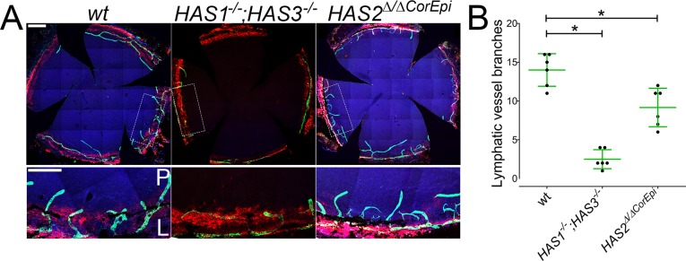
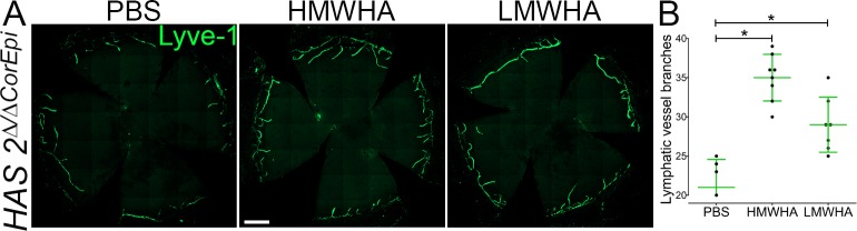
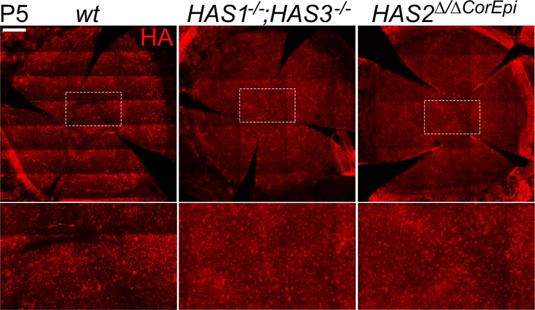
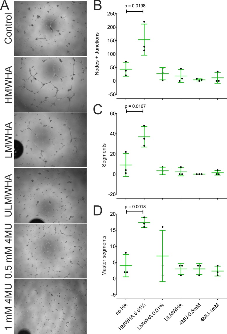
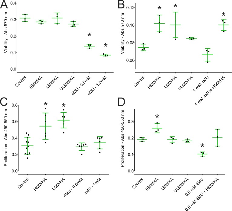
Purpose: We recently reported that the glycosaminoglycan hyaluronan (HA), which promotes inflammatory angiogenesis in other vascular beds, is an abundant component of the limbal extracellular matrix. Consequently, we have explored the possibility that HA contributes to lymphangiogenesis in the inflamed cornea.

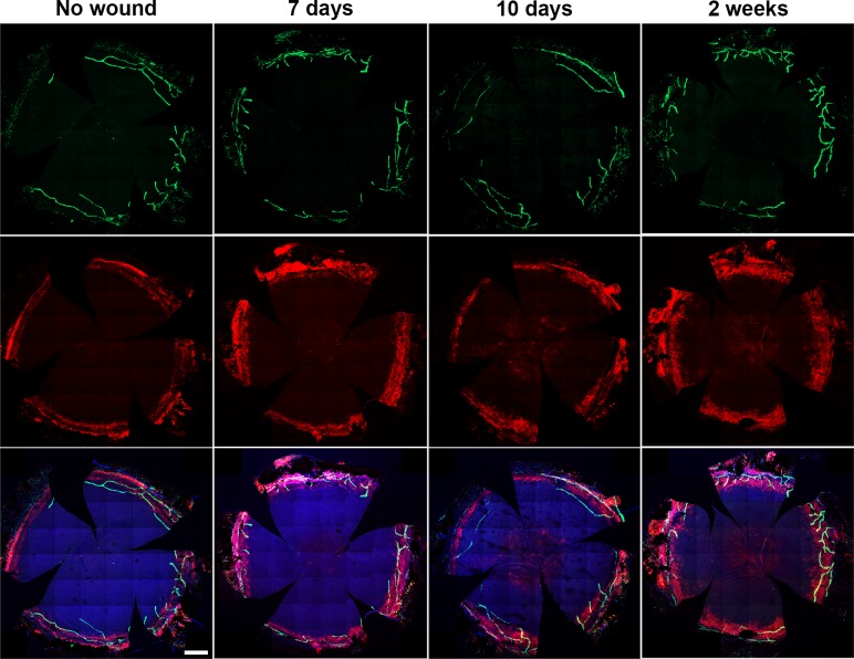
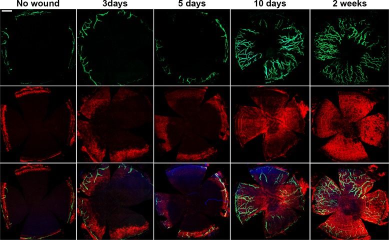
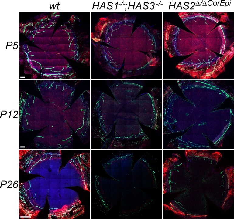
Methods: To study the role of HA on lymphangiogenesis, we used mice lacking the hyaluronan synthases and injury models that induce lymphangiogenesis.

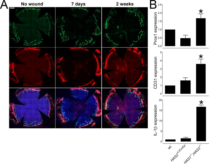
Results: Here we report that HA regulates corneal lymphangiogenesis, both during post-natal development and in response to adult corneal injury. Furthermore, we show that injury to the cornea by alkali burn upregulates both HA production and lymphangiogenesis and that these processes are ablated in HA synthase 2 deficient mice.

Conclusion: These findings raise the possibility that therapeutic blockade of HA-mediated lymphangiogenesis might prevent the corneal scarring and rejection that frequently results from corneal transplantation.


Figures

Figure 1
Figure 1
Lymphatic vessel distribution in wild-type, HAS1−/−;HAS3−/− and HAS2Δ/ΔCorEpi corneas. (A) Corneas were obtained and processed for whole mount staining with anti-LYVE-1 (green) and HABP (red). Zoomed images of the areas demarcated in the top panel, which includes the peripheral cornea (P) and limbal region (L), are shown in the lower panels. Nuclei were counterstained with DAPI (blue). Scale bar represents 500 μm. n = 15. (B) The number of lymphatic vessel branches ≥150 μm was quantified in wild-type, HAS1−/−;HAS3−/− and HAS2Δ/ΔCorEpi corneas by two independent investigators in a blinded manner.
Figure 2
Figure 2
Induction of lymphangiogenesis by alkali burn in wild-type mice. Wild-type mice were subjected to alkali burn and corneas obtained 7 and 10 days and 2 weeks after alkali burn. Uninjured corneas were used as controls. Corneas were obtained and processed for whole mount staining with anti-LYVE-1 (green) and HABP (red). Nuclei were counterstained with DAPI (blue). Scale bar: 500 μm. n = 15.
Figure 3
Figure 3
Induction of lymphangiogenesis by alkali burn in HAS1−/−;HAS3−/− mice. HAS1−/−;HAS3−/− mice were subjected to alkali burn and corneas obtained 3, 5, 7, and 10 days and 2 weeks after alkali burn. Uninjured corneas were used as controls. Corneas were obtained and processed for whole mount staining with anti-LYVE-1 (green) and HABP (red). Nuclei were counterstained with DAPI (blue). Scale bar: 500 μm. n = 15.
Figure 4
Figure 4
Induction of lymphangiogenesis by alkali burn in HAS2Δ/ΔCorEpi mice. (A) HAS2Δ/ΔCorEpi mice were subjected to alkali burn and corneas obtained 7 days and 2 weeks after alkali burn. Uninjured corneas were used as controls. Corneas were obtained and processed for whole mount staining with anti-LYVE-1 (green) and HABP (red). Nuclei were counterstained with DAPI (blue). Scale bar: 500 μm. n = 15. (B) the expression profile of Prox1, CD31, and IL-1β were analyzed 1 week after alkali burn in HAS1−/−;HAS3−/−, HAS2Δ/ΔCorEpi and wild-type mice. n ≥ 5 and asterisk represents P ≤ 0.05.
Figure 5
Figure 5
Effect of HA on lymphangiogenesis in HAS2Δ/ΔCorEpi mice. (A) HA was administered by intrastromal injection into the corneas of HAS2Δ/ΔCorEpi mice to verify whether HA alone can trigger lymphangiogenesis. The distribution of lymphatic vessels was analyzed 10 days after administration. Corneas were obtained and processed for whole mount staining with anti-LYVE-1 (green). Nuclei were counterstained with DAPI (blue). Scale bar: 500 μm. (B) The number of lymphatic vessel branches after PBS, HMWHA, and LMWHA administration were counted in a double-blinded manner by two independent investigators. N = at least 6 and * represents P ≤ 0.05. Data represent the mean ± SD of the total number for each condition.
Figure 6
Figure 6
HA and lymphatic vessel distribution in corneas of mice at post-natal days 5, 12, and 26. The distribution of lymphatic vessels was analyzed in wild-type, HAS1−/−;HAS3−/− and HAS2Δ/ΔCorEpi corneas at post-natal days 5, 12, and 26 (P5, 12 and 26). Corneas were obtained and processed for whole mount staining with anti-LYVE-1 (green) and HABP (red). Nuclei were counterstained with DAPI (blue). Scale bar for P5 and P12 represents 200 μm. Scale bar for P26 represents 500 μm. n = 6.
Figure 7
Figure 7
HA distribution in corneas of mice at post-natal day 5. Corneas were obtained and processed for whole mount staining with HABP (red). The area demarcated by a white dashed box is represented under higher magnification in the lower panel. Scale bar: 500 μm.
Figure 8
Figure 8
Effect of HA and 4-MU on the formation of “tube-like” structures by hDLECs. (A) The tube formation assay was used to study the role of HA on lymphatic vessel formation. The hDLECs were seeded on Matrigel and maintained in the presence or absence of HA or 4-MU. Representative images were acquired of control (untreated), HMWHA, LMWHA, and 1 mM 4-MU-treated hDLECs after 24 hours. (B–D) The ability of the hDLECs to assemble into “tube-like” structures was quantified using an imageJ plugin. The quantitative data for number of nodes, junctions, and segments are represented graphically. The experiment was carried out three times in triplicate, and a mean of each experiment is represented in the graph. The mean ± SD of the total number for each condition is represented in green.
Figure 9
Figure 9
Effect of HA on the viability and proliferation of hDLECs and hLLECs. The MTT assay was used to determine the effect of HA on the viability of hDLECs (A) and hLLECs (B). Human LECs were incubated for 24 hours in the presence or absence of 0.05% HMWHA, 0.05% LMWHA, 0.05% ULMWHA, 0.5 mM 4-MU or 1 mM 4-MU. Values are expressed as mean ± SD of the optical density (OD) measured at 570 nm. BrdU incorporation was used to determine the effect of HA on the proliferation of hDLECs (C) and hLLECs (D). LECs were incubated for 24 hours in the presence or absence of 0.05% HMWHA, 0.05% LMWHA, 0.05% ULMWHA, 0.5 mM 4-MU, or 1 mM 4-MU. Thereafter, BrdU was added and the cells incubated for a further 6 hours. BrdU incorporation was detected using the BrdU Cell Proliferation Assay Kit. The mean ± SD of the total number for each condition is represented in green. Asterisks represents P ≤ 0.05.

Similar articles

Cited by

References

    1. Brown P. Lymphatic system: unlocking the drains. Nature. 2005;436:456–458. - PubMed
    1. Schulte-Merker S, Sabine A, Petrova TV. Lymphatic vascular morphogenesis in development, physiology, and disease. J Cell Biol. 2011;193:607–618. - PMC - PubMed
    1. Hos D, Bachmann B, Bock F, Onderka J, Cursiefen C. Age-related changes in murine limbal lymphatic vessels and corneal lymphangiogenesis. Exp Eye Res. 2008;87:427–432. - PubMed
    1. Chauhan SK, Dohlman TH, Dana R. Corneal lymphatics: role in ocular inflammation as inducer and responder of adaptive immunity. J Clin Cell Immunol. 2014;5(1000256) - PMC - PubMed
    1. Van Buskirk EM. The anatomy of the limbus. Eye (Lond) 1989;3(Pt 2):101–108. - PubMed

Publication types