

Poly-ε-Caprolactone/Gelatin Hybrid Electrospun Composite Nanofibrous Mats Containing Ultrasound Assisted Herbal Extract: Antimicrobial and Cell Proliferation Study
- PMID: 30897714
- PMCID: PMC6474082
- DOI: 10.3390/nano9030462
Poly-ε-Caprolactone/Gelatin Hybrid Electrospun Composite Nanofibrous Mats Containing Ultrasound Assisted Herbal Extract: Antimicrobial and Cell Proliferation Study
Abstract
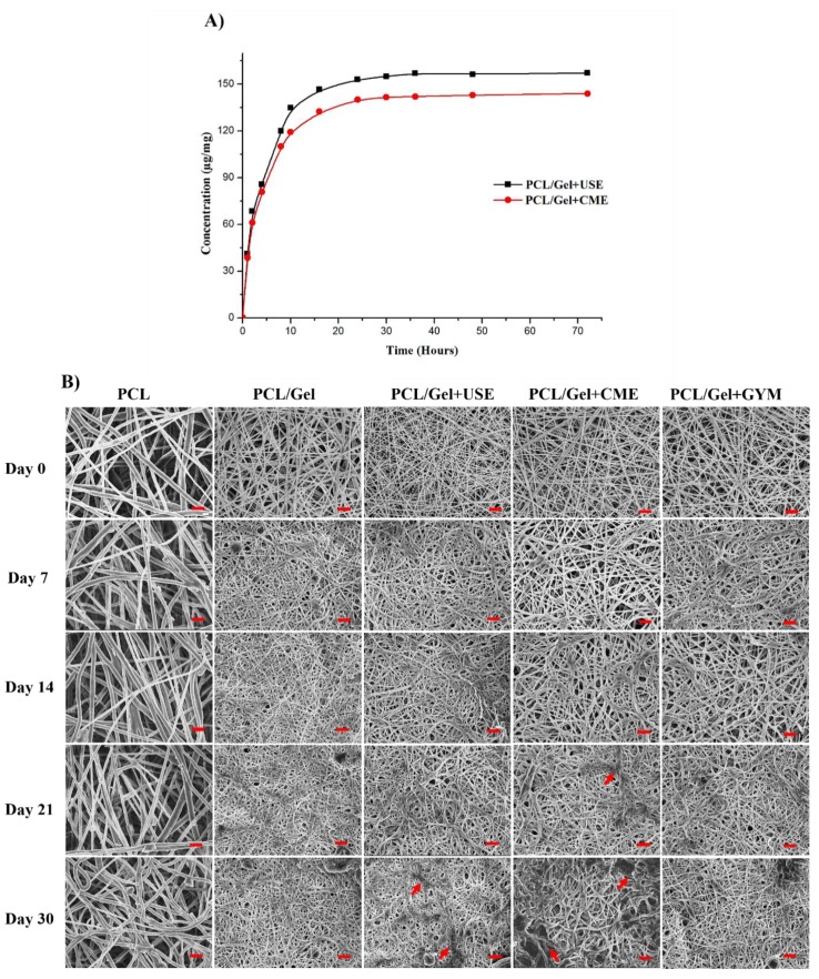
Electrospun fibers have emerged as promising materials in the field of biomedicine, due to their superior physical and cell supportive properties. In particular, electrospun mats are being developed for advanced wound dressing applications. Such applications require the firers to possess excellent antimicrobial properties in order to inhibit potential microbial colonization from resident and non-resident bacteria. In this study, we have developed Poly-ε-Caprolactone /gelatin hybrid composite mats loaded with natural herbal extract (Gymnema sylvestre) to prevent bacterial colonization. As-spun scaffolds exhibited good wettability and desirable mechanical properties retaining their fibrous structure after immersing them in phosphate buffered saline (pH 7.2) for up to 30 days. The initial burst release of Gymnema sylvestre prevented the colonization of bacteria as confirmed by the radial disc diffusion assay. Furthermore, the electrospun mats promoted cellular attachment, spreading and proliferation of human primary dermal fibroblasts and cultured keratinocytes, which are crucial parenchymal cell-types involved in the skin recovery process. Overall these results demonstrated the utility of Gymnema sylvestre impregnated electrospun PCL/Gelatin nanofibrous mats as an effective antimicrobial wound dressing.
Keywords: anti-infective wound dressing; electrospun hybrid mats; gelatin; poly-ε-caprolactone; ultrasound assisted extraction.
Conflict of interest statement
The authors declare no conflict of interest.
Figures


Similar articles
-
Biocompatible Aloe vera and Tetracycline Hydrochloride Loaded Hybrid Nanofibrous Scaffolds for Skin Tissue Engineering.Int J Mol Sci. 2019 Oct 18;20(20):5174. doi: 10.3390/ijms20205174. Int J Mol Sci. 2019. PMID: 31635374 Free PMC article.
-
Antimicrobial properties and biocompatibility of electrospun poly-ε-caprolactone fibrous mats containing Gymnema sylvestre leaf extract.Mater Sci Eng C Mater Biol Appl. 2019 May;98:503-514. doi: 10.1016/j.msec.2018.12.135. Epub 2019 Jan 3. Mater Sci Eng C Mater Biol Appl. 2019. PMID: 30813052
-
Core-Shell Structured Antimicrobial Nanofiber Dressings Containing Herbal Extract and Antibiotics Combination for the Prevention of Biofilms and Promotion of Cutaneous Wound Healing.ACS Appl Mater Interfaces. 2021 Jun 2;13(21):24356-24369. doi: 10.1021/acsami.0c20642. Epub 2021 May 23. ACS Appl Mater Interfaces. 2021. PMID: 34024104
-
Palmatine-loaded electrospun poly(ε-caprolactone)/gelatin nanofibrous scaffolds accelerate wound healing and inhibit hypertrophic scar formation in a rabbit ear model.J Biomater Appl. 2021 Feb;35(7):869-886. doi: 10.1177/0885328220950060. Epub 2020 Aug 16. J Biomater Appl. 2021. PMID: 32799702
-
An Overview on Application of Natural Substances Incorporated with Electrospun Nanofibrous Scaffolds to Development of Innovative Wound Dressings.Mini Rev Med Chem. 2018 Feb 14;18(5):414-427. doi: 10.2174/1389557517666170308112147. Mini Rev Med Chem. 2018. PMID: 28271816 Review.
Cited by
-
Electrospun Sulfonatocalix[4]arene Loaded Blended Nanofibers: Process Optimization and In Vitro Studies.Pharmaceutics. 2022 Sep 9;14(9):1912. doi: 10.3390/pharmaceutics14091912. Pharmaceutics. 2022. PMID: 36145660 Free PMC article.
-
Biocompatible Aloe vera and Tetracycline Hydrochloride Loaded Hybrid Nanofibrous Scaffolds for Skin Tissue Engineering.Int J Mol Sci. 2019 Oct 18;20(20):5174. doi: 10.3390/ijms20205174. Int J Mol Sci. 2019. PMID: 31635374 Free PMC article.
-
Chain-End Functionalization of Poly(ε-caprolactone) for Chemical Binding with Gelatin: Binary Electrospun Scaffolds with Improved Physico-Mechanical Characteristics and Cell Adhesive Properties.Polymers (Basel). 2022 Oct 7;14(19):4203. doi: 10.3390/polym14194203. Polymers (Basel). 2022. PMID: 36236153 Free PMC article.
-
Gelatin Adsorption onto Cellulose Nanocrystals Surfaces at Different pH: A QCM-D Study.Langmuir. 2025 Jun 24;41(24):15319-15330. doi: 10.1021/acs.langmuir.5c00795. Epub 2025 Jun 10. Langmuir. 2025. PMID: 40492952 Free PMC article.
-
Immunomodulation of Macrophages in Diabetic Wound Individuals by Structurally Diverse Bioactive Phytochemicals.Pharmaceuticals (Basel). 2024 Sep 28;17(10):1294. doi: 10.3390/ph17101294. Pharmaceuticals (Basel). 2024. PMID: 39458935 Free PMC article. Review.
References
-
- Rameshbabu A.P., Bankoti K., Datta S., Subramani E., Apoorva A., Ghosh P., Maity P.P., Manchikanti P., Chaudhury K., Dhara S. Silk Sponges Ornamented with Placenta-Derived Extracellular Matrix Augments Full-thickness Cutaneous Wound Healing by Stimulating Neovascularization and Cellular Migration. ACS Appl. Mater. Interfaces. 2018;10:16977–16991. doi: 10.1021/acsami.7b19007. - DOI - PubMed
-
- Baranowska-Korczyc A., Warowicka A., Jasiurkowska-Delaporte M., Grześkowiak B., Jarek M., Maciejewska B.M., Jurga-Stopa J., Jurga S. Antimicrobial electrospun poly(ε-caprolactone) scaffolds for gingival fibroblast growth. RSC Adv. 2016;6:19647–19656. doi: 10.1039/C6RA02486F. - DOI
Grants and funding
- CSR-KN/CRS-70/2015-16/811/UGC-DAE Consortium for Scientific Research, University Grants Commission
- NMRC/TCR/008-SERI/2013/National Research Foundation Singapore
- NMRC/CBRG/0048/2013/Singapore National Medical Research Council
- . R1181/83/2014/SNEC Ophthalmic Technologies Incubator Program grant
- L0412290/Nanyang Technological University Singapore Start-Up Grant
LinkOut - more resources
Full Text Sources
Molecular Biology Databases
Research Materials
