Review
. 2019 Mar 20;8(3):264.
doi: 10.3390/cells8030264.

Cysteine Cathepsins and their Extracellular Roles: Shaping the Microenvironment

Affiliations
Review

Cysteine Cathepsins and their Extracellular Roles: Shaping the Microenvironment

Eva Vidak et al. Cells. .

Abstract

: For a long time, cysteine cathepsins were considered primarily as proteases crucial for nonspecific bulk proteolysis in the endolysosomal system. However, this view has dramatically changed, and cathepsins are now considered key players in many important physiological processes, including in diseases like cancer, rheumatoid arthritis, and various inflammatory diseases. Cathepsins are emerging as important players in the extracellular space, and the paradigm is shifting from the degrading enzymes to the enzymes that can also specifically modify extracellular proteins. In pathological conditions, the activity of cathepsins is often dysregulated, resulting in their overexpression and secretion into the extracellular space. This is typically observed in cancer and inflammation, and cathepsins are therefore considered valuable diagnostic and therapeutic targets. In particular, the investigation of limited proteolysis by cathepsins in the extracellular space is opening numerous possibilities for future break-through discoveries. In this review, we highlight the most important findings that establish cysteine cathepsins as important players in the extracellular space and discuss their roles that reach beyond processing and degradation of extracellular matrix (ECM) components. In addition, we discuss the recent developments in cathepsin research and the new possibilities that are opening in translational medicine.

Keywords: cancer; cathepsin; extracellular matrix; inflammation associated disease; osteoporosis.


Conflict of interest statement

The authors declare no conflict of interest.

Figures

Figure 1
Figure 1
Cysteine cathepsin structures and specificities. (A) Crystal structure of the two-chain form of cathepsin L (PDB 1icf [33]) in standard orientation colored according to surface hydrophobicity (red: most hydrophobic, blue: most hydrophilic). Active site Cys25 is colored in yellow. (B) Substrate specificity of different cysteine cathepsins relative to the cleavage site, which is between the P1 and P1’ residues. S4–S1 and S1'–S2' represent the substrate binding sites into which the corresponding substrate residues P4–P2’ bind, with P4–P1 designating the substrate residue N-terminals of the cleavage site and P1’–P2’ designating residue C-terminals of the cleavage site, respectively. Amino acid residues of the substrate, which bind to their designated sites, are shown with colored circles, with each color representing a different amino acid class required for the binding of the substrate to the active site.
Figure 2
Figure 2
Cysteine cathepsin secretion and their extracellular roles. (A) Secretion of extracellular cathepsin is often tightly connected with their overexpression and can be triggered by diverse cell signaling pathways. Overexpressed cysteine cathepsins are usually secreted with vesicular exocytosis. (B) In the extracellular milieu, cysteine cathepsins cleave different targets. Cleavages of cell adhesion molecules (CAM), cell-cell contacts, and proteins of ECM mainly influence cell adhesion and migration. Additionally, proteolytic products of these cleavages can act as signaling molecules and have an impact on cell growth, invasion, and angiogenesis. Other main target of cysteine cathepsins are cell receptors, and their cleavage can result in either constantly triggered signaling, in the case of partial trimming of the receptor, or inhibited signaling, in the case of a complete removal of the extracellular domain. CAM, cell adhesion molecules; ECM, extracellular matrix; IL, interleukin; RANK, receptor activator of NF-κΒ; RANKL, receptor activator of NF-κΒ ligand; STAT, signal transducer and activator of transcription; TFEB, transcription factor EB; TNFα, tumor necrosis factor alpha; TNFR1, tumor necrosis factor receptor 1.
Figure 3
Figure 3
Extracellular cathepsins as diagnostic targets, prodrug activators, and targets for targeted drug delivery. The high levels of cathepsins in the ECM can be utilized in different imaging and targeting techniques. (A) Fluorescent substrates, substrate-based probes, and activity-based probes are the most commonly used tools for in vivo and in vitro imaging. (B) Extracellular cathepsins can be also used for targeted drug delivery and for prodrug activation. (C) Since many cathepsins are also overexpressed and active in different cancers, they can be targeted directly using their inhibitors, designed ankyrin repeat proteins (DARPins), or antibodies. These molecules can also be conjugated and, as such, used for drug delivery and tumor visualization.

Similar articles

Cited by

References

    1. Turk V., Stoka V., Vasiljeva O., Renko M., Sun T., Turk B., Turk D. Cysteine cathepsins: From structure, function and regulation to new frontiers. Biochim. Biophys. Acta. 2012;1824:68–88. doi: 10.1016/j.bbapap.2011.10.002. - DOI - PMC - PubMed
    1. Turk V., Turk B., Turk D. Lysosomal cysteine proteases: Facts and opportunities. EMBO J. 2001;20:4629–4633. doi: 10.1093/emboj/20.17.4629. - DOI - PMC - PubMed
    1. Rossi A., Deveraux Q., Turk B., Sali A. Comprehensive search for cysteine cathepsins in the human genome. Biol. Chem. 2004;385:363–372. doi: 10.1515/BC.2004.040. - DOI - PubMed
    1. Vidmar R., Vizovisek M., Turk D., Turk B., Fonovic M. Protease cleavage site fingerprinting by label-free in-gel degradomics reveals pH-dependent specificity switch of legumain. EMBO J. 2017;36:2455–2465. doi: 10.15252/embj.201796750. - DOI - PMC - PubMed
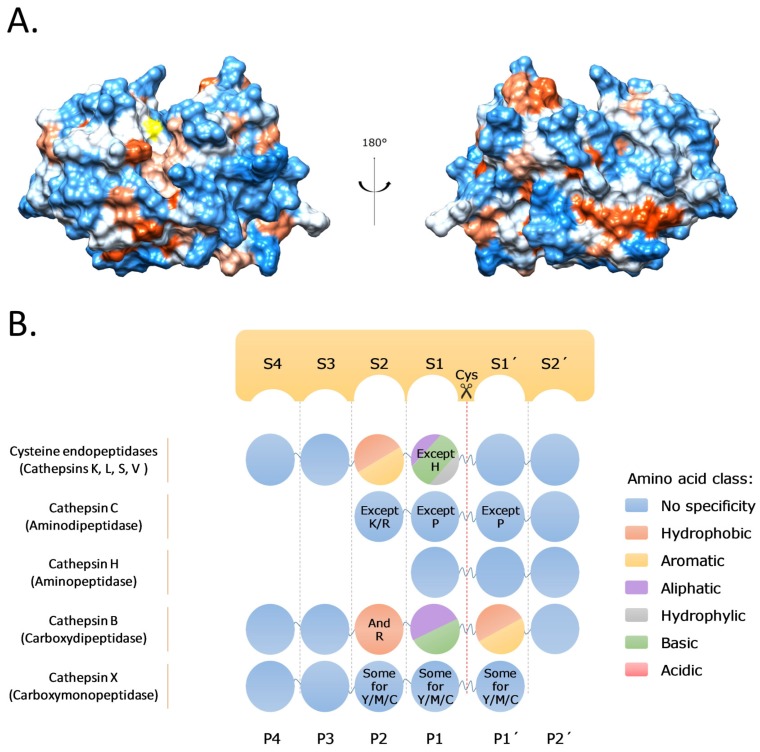
    1. Vizovisek M., Vidmar R., Van Quickelberghe E., Impens F., Andjelkovic U., Sobotic B., Stoka V., Gevaert K., Turk B., Fonovic M. Fast profiling of protease specificity reveals similar substrate specificities for cathepsins K, L and S. Proteomics. 2015;15:2479–2490. doi: 10.1002/pmic.201400460. - DOI - PubMed

Publication types

LinkOut - more resources