

Intermittent Parathyroid Hormone [1-34] Augments Chondrogenesis of the Mandibular Condylar Cartilage of the Temporomandibular Joint
- PMID: 30897936
- PMCID: PMC8461153
- DOI: 10.1177/1947603519833146
Intermittent Parathyroid Hormone [1-34] Augments Chondrogenesis of the Mandibular Condylar Cartilage of the Temporomandibular Joint
Abstract
Objective: To characterize the long-term effects of intermittent parathyroid hormone (I-PTH) on the mandibular condylar cartilage (MCC) and subchondral bone of the temporomandibular joint, in vivo and in vitro.
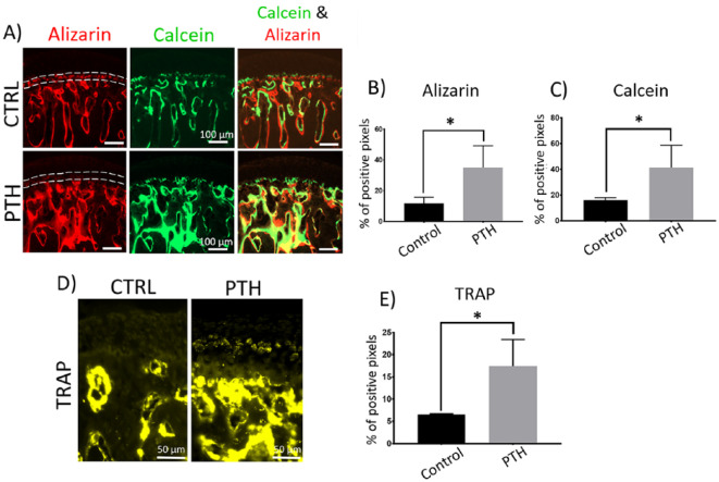
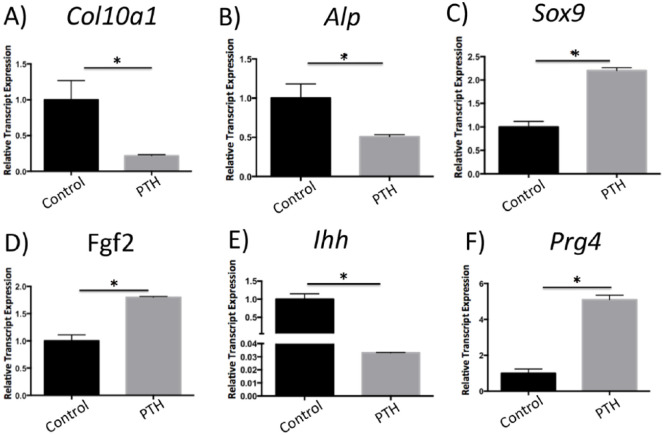
Materials and methods: For the in vivo experiments, sixteen 10-week-old mice were divided into 2 groups: (1) I-PTH (n = 8)-subcutaneous daily injection of PTH; (2) control group (n = 8)-subcutaneous daily injection of saline solution. Experiments were carried out for 4 weeks. Mice were injected with calcein, alizarin complexone, and cell proliferation marker before euthanasia. For the in vitro experiments, primary chondrocyte cultures from the MCC of eight 10-week-old mice were treated with I-PTH for 14 days.
Results: There was a significant increase in bone volume, tissue density, mineral deposition, osteoclastic activity, cell proliferation in the cartilage, and cartilage thickness in the I-PTH-treated mice when compared with the control group. In addition, immunohistochemistry in cartilage revealed that I-PTH administration led to an increase in expression of vascular endothelial growth factor and to a decreased expression of sclerostin, matrix metallopeptidase 13, and aggreganase-1 (ADAM-TS4). Quantitative polymerase chain reaction analysis of the I-PTH-treated chondrocytes revealed significantly decreased relative expression of collagen type X (Col10a1), alkaline phosphatase (Alp), and Indian Hedgehog (Ihh) and remarkable increased expression of Sox9, fibroblast growth factor 2 (Fgf2), and proteoglycan 4 (Prg4).
Conclusion: I-PTH administration causes anabolic effects at the subchondral region of the mandibular condyle while triggers anabolic and protective effects at the MCC.
Keywords: mandibular condylar cartilage; parathyroid hormone; temporomandibular joint.
Conflict of interest statement
Figures


Similar articles
-
Anabolic Response of Intermittent Parathyroid Hormone and Alendronate on the Osteochondral Tissue of TMJ.Cartilage. 2022 Dec;13(4):171-183. doi: 10.1177/19476035221109229. Epub 2022 Oct 14. Cartilage. 2022. PMID: 36239576 Free PMC article.
-
PTH [1-34]-induced alterations predispose the mandibular condylar cartilage to mineralization.Orthod Craniofac Res. 2017 Jun;20 Suppl 1:162-166. doi: 10.1111/ocr.12157. Orthod Craniofac Res. 2017. PMID: 28643904
-
PTH [1-34] induced differentiation and mineralization of mandibular condylar cartilage.Sci Rep. 2017 Jun 12;7(1):3226. doi: 10.1038/s41598-017-03428-y. Sci Rep. 2017. PMID: 28607469 Free PMC article.
-
Mandibular Condylar Cartilage in Development and Diseases: A PTHrP-Centric View.Orthod Craniofac Res. 2025 Apr 19. doi: 10.1111/ocr.12936. Online ahead of print. Orthod Craniofac Res. 2025. PMID: 40251915 Review.
-
The Roles of Indian Hedgehog Signaling in TMJ Formation.Int J Mol Sci. 2019 Dec 13;20(24):6300. doi: 10.3390/ijms20246300. Int J Mol Sci. 2019. PMID: 31847127 Free PMC article. Review.
Cited by
-
Anabolic Response of Intermittent Parathyroid Hormone and Alendronate on the Osteochondral Tissue of TMJ.Cartilage. 2022 Dec;13(4):171-183. doi: 10.1177/19476035221109229. Epub 2022 Oct 14. Cartilage. 2022. PMID: 36239576 Free PMC article.
-
Parathyroid hormone promotes cartilage healing after free reduction of mandibular condylar fractures by upregulating Sox9.Exp Biol Med (Maywood). 2021 Nov;246(21):2249-2258. doi: 10.1177/15353702211027114. Epub 2021 Jul 7. Exp Biol Med (Maywood). 2021. PMID: 34233524 Free PMC article.
References
-
- Hansen S, Hauge EM, Jensen JEB, Brixen K. Differing effects of PTH 1-34, PTH 1-84, and zoledronic acid on bone microarchitecture and estimated strength in postmenopausal women with osteoporosis: an 18-month open-labeled observational study using HR-pQCT. J Bone Miner Res. 2013;28(4):736-45. - PubMed
-
- Yamazaki K, Suda N, Kuroda T.Distribution of parathyroid hormone-related protein (PTHrP) and type I parathyroid hormone (PTH) PTHrP receptor in developing mouse mandibular condylar cartilage. Arch Oral Biol. 1999;44(10):853-60. - PubMed
-
- Orth P, Cucchiarini M, Zurakowski D, Menger MD, Kohn DM, Madry H.Parathyroid hormone [1-34] improves articular cartilage surface architecture and integration and subchondral bone reconstitution in osteochondral defects in vivo. Osteoarthritis Cartilage. 2013;21(4):614-24. - PubMed
Publication types
MeSH terms
Substances
Grants and funding
LinkOut - more resources
Full Text Sources
Research Materials
Miscellaneous
