. 2019;8(1):438-447.
doi: 10.1080/22221751.2019.1590128.

Replication and virulence in pigs of the first African swine fever virus isolated in China

Affiliations

Replication and virulence in pigs of the first African swine fever virus isolated in China

Dongming Zhao et al. Emerg Microbes Infect. 2019.

Abstract

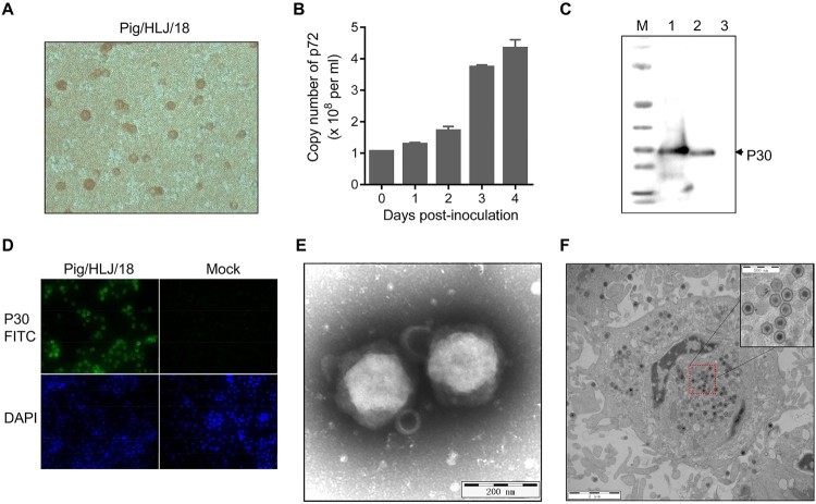
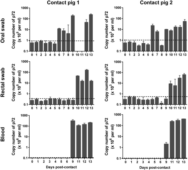
African swine fever (ASF) entered China in August 2018 and rapidly spread across the entire country, severely threatening the Chinese domestic pig population, which accounts for more than 50% of the pig population worldwide. In this study, an ASFV isolate, Pig/Heilongjiang/2018 (Pig/HLJ/18), was isolated in primary porcine alveolar macrophages (PAMs) from a pig sample from an ASF outbreak farm. The isolate was characterized by using the haemadsorption (HAD) test, Western blotting and immunofluorescence, and electronic microscopy. Phylogenetic analysis of the viral p72 gene revealed that Pig/HLJ/18 belongs to Genotype II. Infectious titres of virus propagated in primary PAMs and pig marrow macrophages were as high as 107.2 HAD50/ml. Specific-pathogen-free pigs intramuscularly inoculated with different virus dosages at 103.5-106.5 HAD50 showed acute disease with fever and haemorrhagic signs. The incubation periods were 3-5 days for virus-inoculated pigs and 9 days for contact pigs. All virus-inoculated pigs died between 6-9 days post-inoculation (p.i.), and the contact pigs died between 13-14 days post-contact (p.c.). Viremia started on day 2 p.i. in inoculated pigs and on day 9 p.c. in contact pigs. Viral genomic DNA started to be detected from oral and rectal swab samples on 2-5 days p.i. in virus-inoculated pigs, and 6-10 days p.c. in contact pigs. These results indicate that Pig/HLJ/18 is highly virulent and transmissible in domestic pigs. Our study demonstrates the threat of ASFV and emphasizes the need to control and eradicate ASF in China.

Keywords: African swine fever virus; animal study; pig; transmission; virus isolation.


Figures

Figure 1.
Figure 1.
Detection of ASFV in a field sample of spleen tissue. (A) PCR detection of spleen DNA from the suspected pig. Lane 1, sample; lane 2, positive control plasmid; lane 3, negative control. (B) HAD assay of the spleen homogenate. The 10-times dilution of the supernatant of the homogenate was inoculated into pig PBMCs with 1% pig blood cells. HAD was observed for 7 days.
Figure 2.
Figure 2.
Viral growth in primary PAMs. Primary PAMs were infected with Pig/HLJ/18 at an MOI of 0.2, and the supernatants were collected for infectious virus particle detection and quantification of viral p72 gene copies by using HAD (A) and qPCR (B) assays, respectively. The expression of viral p30 in the infected primary PAMs was detected by Western blotting of the lysate of cells infected with Pig/HLJ/18 (C, line 1) [cells transfected with pCAGG-p30 (C, line 2)] and by use of an immunofluorescence assay with cells infected with Pig/HLJ/18 (D). Electron microscopic observation of ASFV particles in cell culture supernatants by negative staining (E), and in infected cells by ultrathin-sectioning (F) at 48 h post-infection.
Figure 3.
Figure 3.
Phylogenetic analysis of Pig/HLJ/18 based on its partial p72 gene. The sequences of the p72 genes of representative ASFVs were downloaded from the NCBI database. The neighbor-joining method was used to construct phylogenetic trees using MEGA X software (https://www.megasoftware.net/). Numbers along branches indicate bootstrap values >70% (1,000 replicates). The black diamond indicates the ASFV isolate from this study. The white triangle marks the ASFV sequences from the first case in China. Scale bars indicate nucleotide substitutions per site.
Figure 4.
Figure 4.
Viral genomic copies in samples collected from Pig/HLJ/18 infected pigs. Blood, as well as oral and rectal swab samples, were collected at the indicated time points from three pigs inoculated with 103.5 HAD50 of Pig/HLJ/18. Viral p72 gene copies were quantified by using the qPCR assay. The dashed black lines in these panels indicate the limit of detection compared with the data of samples collected at day 0 post-inoculation.
Figure 5.
Figure 5.
Viral titres in the blood and organs of pigs tested by using the HAD assay. Blood was collected from pigs that were inoculated with different doses of ASFV Pig/HLJ/18 (A─D) or from pigs that contacted with pigs that were inoculated with ASFV Pig/HLJ/18 (E─F) at the indicated time points. (A) 106.5 HAD50-inoculated pig; (B) 105.5 HAD50-inoculated pigs; (C) 104.5 HAD50-inoculated pigs; (D) 103.5 HAD50-inoculated pigs; (E) Pig co-housed with pigs inoculated with 104.5 HAD50 of ASFV Pig/HLJ/18; (F) Pig co-housed with pigs inoculated with 103.5 HAD50 of ASFV Pig/HLJ/18. (G) Viral titres in the liver and spleen from virus-inoculated pigs and contact pigs. Samples were collected during necropsy and the virus inoculation dose for each pig group is shown at the top of the panel.
Figure 6.
Figure 6.
Viral genomic copies in samples from contact pigs. Blood, as well as oral and rectal swab samples were collected from two contact pigs, which were housed with pigs inoculated with 104.5 HAD50 (contact pig 1) and 103.5 HAD50 (contact pig 2), respectively, at different timepoints. The dashed black lines in these panels indicate the limit of detection compared with the data of samples collected at day 0 post-inoculation.
Figure 7.
Figure 7.
Gross lesions in pigs that died as a result of infection with Pig/HLJ/18. The gross lesions of all pigs are summarized and shown in Supplementary Table. The lesions of the heart, lung, gall bladder, spleen, mesenteric lymph nodes, and gastro-hepatic lymph nodes of the pig that was inoculated with 106.5 HAD50 of the virus are shown here. A. Severe and diffuse haemorrhage of the epicardium in the heart. B. Mild interstitial oedema of the lung; the insert is a higher magnification of the framed area. C. Severe haemorrhage and oedema of the gallbladder wall and liver; the insert is a higher magnification of the circled area. D. Severe, enlarged, friable, and dark black appearance of the spleen. E. Mesenteric lymph nodes, showing mildly enlarged and red-black appearance. F. Gastro-hepatic lymph nodes appear very enlarged and haemorrhagic.

Similar articles

Cited by

References

    1. De la Torre A, Bosch J, Iglesias I, et al. . Assessing the risk of African swine fever introduction into the European Union by wild boar. Transbound Emerg Dis. 2015;62:272–279. doi: 10.1111/tbed.12129 - DOI - PubMed
    1. Dixon LK, Chapman DA, Netherton CL, et al. . African swine fever virus replication and genomics. Virus Res. 2013;173:3–14. doi: 10.1016/j.virusres.2012.10.020 - DOI - PubMed
    1. Revilla Y, Perez-Nunez D, Richt JA.. African swine fever virus biology and vaccine approaches. Adv Virus Res. 2018;100:41–74. doi: 10.1016/bs.aivir.2017.10.002 - DOI - PubMed
    1. Galindo I, Alonso C.. African swine fever virus: A review. Viruses. 2017;9:103. doi: 10.3390/v9050103 - DOI - PMC - PubMed
    1. Arzt J, White WR, Thomsen BV, et al. . Agricultural diseases on the move early in the third millennium. Vet Pathol. 2010;47:15–27. doi: 10.1177/0300985809354350 - DOI - PubMed