. 2019 Mar 21;38(1):134.
doi: 10.1186/s13046-019-1130-2.

Discovery of a natural small-molecule compound that suppresses tumor EMT, stemness and metastasis by inhibiting TGFβ/BMP signaling in triple-negative breast cancer

Affiliations

Discovery of a natural small-molecule compound that suppresses tumor EMT, stemness and metastasis by inhibiting TGFβ/BMP signaling in triple-negative breast cancer

Lei Di et al. J Exp Clin Cancer Res. .

Abstract

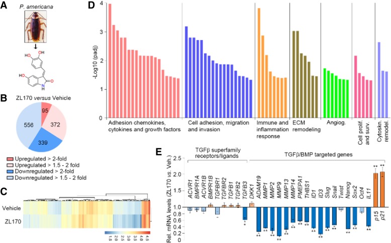
Background: The transforming growth factor β (TGFβ) and bone morphogenetic protein (BMP) signaling pathways are both constitutively activated in triple-negative breast cancer (TNBC). We are interested in isolating the naturally-derived small-molecule inhibitor that could simultaneously targeting TGFβ/BMP pathways and further studying its anti-proliferative/-metastatic effects as well as the underlying mechanisms in multiple tumor models.

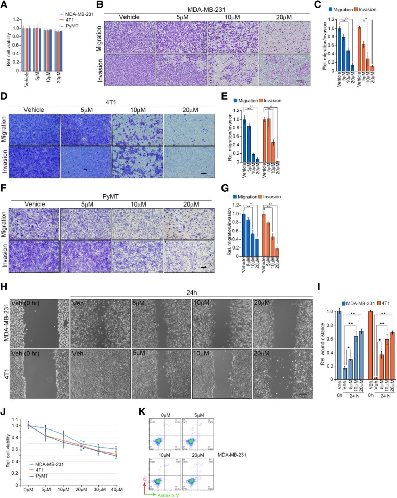
Methods: Multiple in vitro cell-based assays are used to examine the compound's inhibitory efficacy on TNBC cell growth, stemness, epithelial-mesenchymal transition (EMT), invasion and migration by targeting TGFβ/BMP signaling pathways. Transgenic breast cancer mouse model (MMTV-PyMT), subcutaneous xenograft and bone metastasis models are used to examine ZL170's effects on TNBC growth and metastasis potentials in vivo.

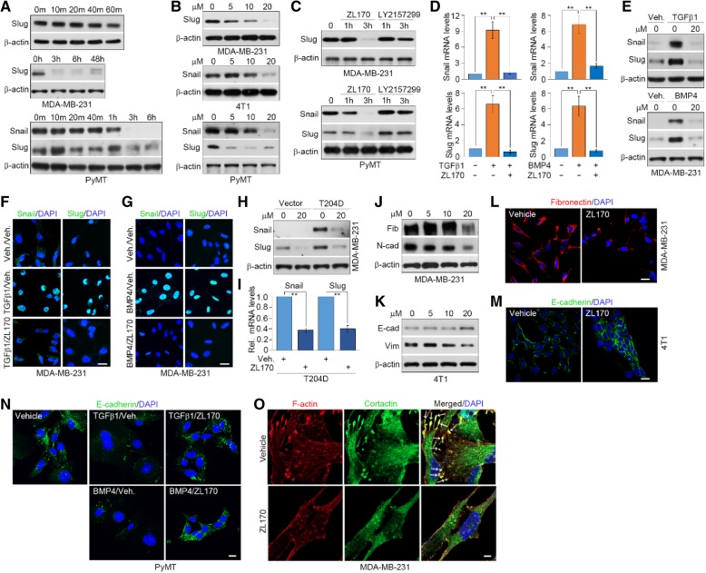
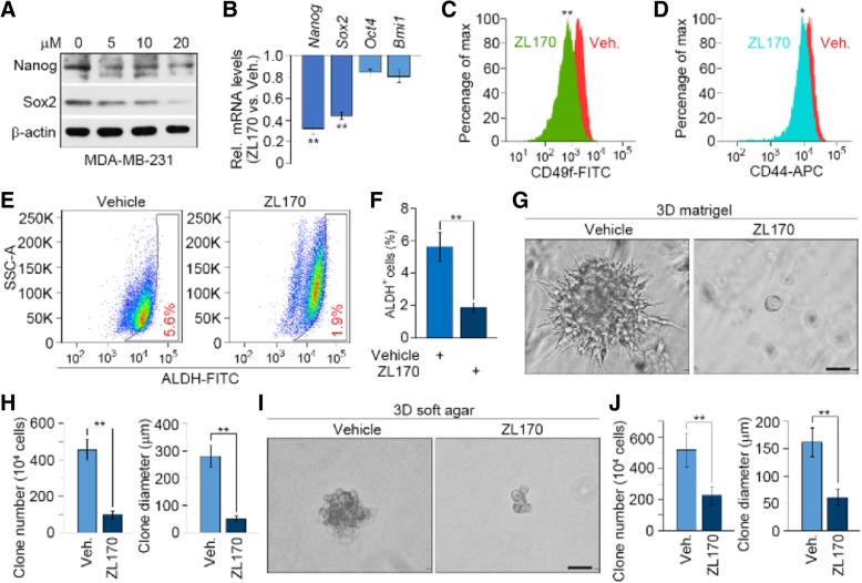
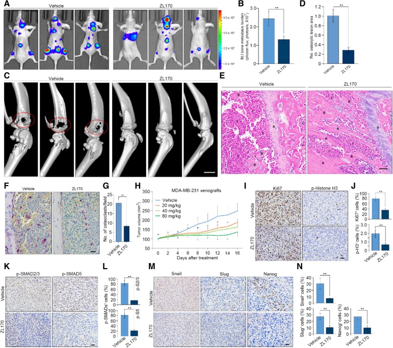
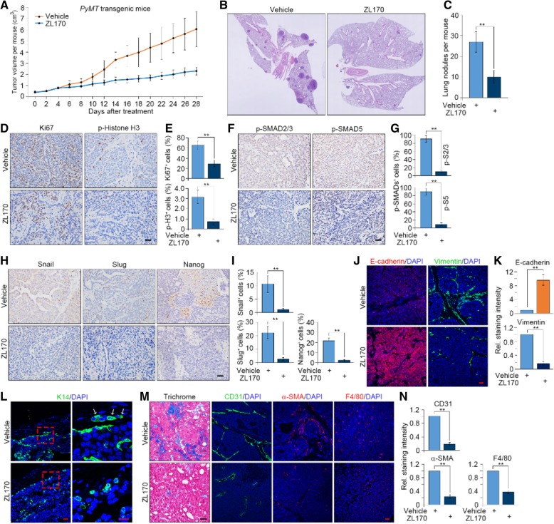
Results: ZL170 dose-dependently inhibits cell proliferation, EMT, stemness, invasion and migration in vitro via specifically targeting canonical TGFβ/BMP-SMADs pathways in TNBC cells. The compound significantly hinders osteolytic bone metastasis and xenograft tumor growth without inflicting toxicity on vital organs of tumor-bearing nude mice. ZL170 strongly inhibits primary tumor growth and lung metastases in MMTV-PyMT transgenic mice. ZL170-treated tumors exhibit impaired TGFβ/BMP signaling pathways in both epithelial and stromal compartments, thereby creating a suppressive tumor microenvironment characterized by reduced extracellular matrix deposition and decreased infiltration of stromal cells.

Conclusions: ZL170 inhibits tumor EMT, stemness and metastasis and could be further developed as a potent anti-metastatic agent used in combination with cytotoxic drugs for treatment of TNBC and other advanced metastatic cancers.

Keywords: Epithelial–mesenchymal transition; Metastasis; TGFβ/BMP; Triple-negative breast cancer; ZL170.


Conflict of interest statement

Ethics approval and consent to participate

The experimental protocol was approved by the Animal Welfare and Ethics Committee of China Pharmaceutical University.

Consent for publication

Not applicable.

Competing interests

The authors declare that they have no competing interests.

Publisher’s Note

Springer Nature remains neutral with regard to jurisdictional claims in published maps and institutional affiliations.

Figures

Fig. 1
Fig. 1
ZL170 targets the TGFβ and BMP signaling pathways in MDA-MB-231 cells. a Chemical structure of ZL170 that was extracted from the dry whole bodies of P. americana. b-d RNA sequencing analysis of vehicle- and ZL170-treated MDA-MB-231 cells. Pie chart denotes the distribution of total transcripts changed in ZL170-treated cells as compared with vehicle-treated cells (b). Heat map of all targets in vehicle- and ZL170-treated cells (c). Gene set enrichment analysis (GSEA) of expression of signature genes involved in multiple cellular functions (d). e qPCR analysis validated TGFβ/BMP-targeted genes as well as genes encoding TGFβ superfamily receptors and ligands. Data are represented as mean ± S.D. (n = 3 independent experiments). * P < 0.05, ** P < 0.01, two-sided Student’s t-test
Fig. 2
Fig. 2
ZL170 reduces TNBC cell migration, invasion and proliferation, but does not induce apoptosis in vitro. a Cell viability of MDA-MB-231, 4 T1 and PyMT cells treated with vehicle or 20 μM ZL170 for 24 h (n = 3) (b, d and f) Boyden chamber migration and invasion assays of MDA-MB-231 (b), 4 T1 (d) and PyMT (f) cells (n = 3). Cells pretreated with ZL170 at different doses for 24 h were seeded in upper insert in the presence (for invasion assay) or absence (for migration assay) of pre-coated Matrigel. c, e and g Quantification of the migrated or invaded cells as shown in c, e and g, respectively. h Migration wound healing assay of MDA-MB-231 and 4 T1 cells in the presence of different doses of ZL170. i Quantification of the wound healing as shown in h. j Cell viability of MDA-MB-231, 4 T1 and PyMT cells treated with vehicle or different doses of ZL170 for 48 h (n = 3). k Cell apoptosis analysis of MDA-MB-231 cells that were treated with vehicle or different doses of ZL170 for 48 h (n = 3). Cells were co-stained with Annexin V and PI. Data are represented as mean ± S.D. * P < 0.05, ** P < 0.01, one-way ANOVA test. Scale bars, 200 μm (b, d, f, h)
Fig. 3
Fig. 3
ZL170 is a dual inhibitor of TGFβ and BMP kinase receptors and reduces activation of Smads in TNBC cells. a In vitro kinase activity assays of the inhibitory efficacy of ZL170 on phosphorylation of the substrates (BMPR1A, BMPR1B, BMPR2, TGFBR1, ACVR1B, ACVR1 and TGFBR2). b Molecular docking analysis of the potential binding between ZL170 and TGFβ/BMP receptors. Illustration of surface crystal structure of ZL170 against BMPR1A, BMPR1B, BMPR2 and TGFBR1 shown. c Representative immunoblot analyses of the levels of phospho-Smad1/5 and phospho-Smad2/3 (and their total forms) in MDA-MB-231 and PyMT cells that were treated with vehicle or ZL170 at 20 μM for different times (n = 3). d Immunoblot analyses of the levels of phospho-Smad1/5 and phospho-Smad2/3 (and their total forms) in the indicated cells that were treated with vehicle or ZL170 at 5, 10 and 20 μM for 3 h. n = 3 independent experiments. e ZL170 efficiently abolished TGFβ1-stimulated and BMP4-stimulated expression of phospho-Smads in MDA-MB-231 cells as indicated by representative immunoblot analyses (n = 3). Cells were treated with TGFβ1 or BMP4 in combination with ZL170 at 20 μM for 3 h. f and g Representative immunofluorescent staining of phospho-Smads (f) and Smads (g) in resting, TGFβ1-stimulated and BMP4-stimulated MDA-MB-231 cells (n = 3). (H) ZL170 efficiently reduced the levels of phospho-Smads in TGFBR1-T204D (left panel) or BMPR1A-Q233D (right panel) stably expressing cells as indicated by representative immunoblot analyses (n = 3). Cells were treated with the compound for 3 h. i SBE promoter luciferase reporter assays in MDA-MB-231 cells that were treated with TGFβ1 and increasing doses of ZL170 (left panel) or in TGFBR1-T204D stably expressing cells treated with increasing doses of ZL170 (right panel) (n = 3). j BRE4 promoter luciferase reporter assays in MDA-MB-231 cells treated with increasing doses of ZL170 (left panel) or in BMPR1A-Q233D stably expressing cells treated with increasing doses of ZL170 (right panel). k-m Boyden chamber invasion assays of MDA-MB-231 cells stably expressing control-shRNA or TGFBR1-shRNA (k and l). Quantification of invaded cells were shown in (M). n = 3 independent experiments. n-p Boyden chamber invasion assays of MDA-MB-231 cells stably expressing control-shRNA or BMPR1A-shRNA (n and o). Quantification of invaded cells were shown in (p). n = 3 independent experiments. Data are represented as mean ± S.D. * P < 0.05, ** P < 0.01, one-way ANOVA test. Scale bars = 20 μm (f, g) and 200 μm (l, o)
Fig. 4
Fig. 4
ZL170 inhibits Snail and Slug expression, reverses the EMT program and reduces EMT-dependent FLP formation in TNBC cells. a Representative immunoblot analyses of Snail and Slug levels in MDA-MB-231 and PyMT cells that were treated with vehicle or ZL170 at 20 μM for different times (n = 3). b and c Immunoblot analyses of Snail and Slug levels in the indicated cells that were treated with vehicle or ZL170 at 5, 10 and 20 μM for 3 h (b) or in the indicated cells that were treated with vehicle, ZL170 or LY2157299 at 20 μM for 1 h and 3 h (c). n = 3 independent experiments. d-g ZL170 efficiently abolished TGFβ1-stimulated and BMP4-stimulated expression of Snail and Slug in MDA-MB-231 cells as indicated by qPCR (d), immunoblot (e) and immunofluorescent (f and g) analyses (n = 3). Cells were treated with TGFβ1 or BMP4 in combination with ZL170 at 20 μM for 3 h. Data are represented as mean ± S.D. ** P < 0.01, one-way ANOVA test. h and i ZL170 efficiently reduced Snail and Slug levels in TGFBR1-T204D stably expressing cells as indicated by representative immunoblot (h) and qPCR (i) analyses (n = 3). Cells were treated with the compound for 3 h. j and k Representative immunoblot analyses of fibronectin, N-cadherin, vimentin or E-cadherin levels in MDA-MB-231 (j) and 4 T1 (k) cells that were treated with vehicle or ZL170 at 5, 10 and 20 μM for 48 h (n = 3). (L and M) Immunofluorescent staining of fibronectin (l) and E-cadherin (m) in the indicated cells that were treated as described in J and K, respectively (n = 3). n Immunofluorescent staining of E-cadherin in the indicated cells that were treated with TGFβ1 or BMP4 in combination with ZL170 at 20 μM for 48 h (n = 3). o Immunofluorescent staining of F-actin and cortactin in vehicle- and ZL170-treated MDA-MB-231 cells. Nuclear, DAPI (blue). Arrows denote F-actin+/cortactin+ FLPs (n = 3). Scale bars = 20 μm (f, g, l, m, n) and 5 μm (o)
Fig. 5
Fig. 5
ZL170 reduces stemness function in MDA-MB-231 cells. a Immunoblot analyses of Nanog and Sox2 levels in MDA-MB-231 cells that were treated with vehicle or ZL170 at 5, 10 and 20 μM for 48 h (n = 3). b qPCR analysis of Nanog, Sox2, Oct4 and Bmi1 levels in cells that were treated as described in A (n = 3). c and d Flow cytometry analysis of CD49f (c) and CD44 (d) cell-surface expression in MDA-MB-231 cells that were treated with vehicle or ZL170 for 48 h (n = 3). e and f Representative histogram (e) and quantification (f) of ALDH+ subpopulation in MDA-MB-231 cells that were treated with vehicle or ZL170 for 48 h (n = 3). g-j Representative phase-contrast images of vehicle-treated or ZL170-treated MDA-MB-231 cells that were embedded in 3D Matrigel (g) or 3D soft agar gel (i), and tumor sphere number and diameter quantified (h and j). n = 3 independent experiments. Data are represented as mean ± S.D. * P < 0.05, ** P < 0.01, two-sided Student’s t-test. Scale bars = 50 μm (g, i)
Fig. 6
Fig. 6
ZL170 inhibits TNBC osteolytic bone metastasis and xenograft tumor growth by targeting the TGFβ and BMP signaling pathways. a and b BLI images (a) and quantification (b) of bone lesions from nude mice that were intracardially injected with SCP2 cells labelled with firefly luciferase and then received treatment of vehicle or ZL170 (i.p., 80 mg/kg/day for 2 consecutive weeks; n = 6 mice). c and d μ-CT images (c) and quantification (d) of osteolytic lesions from vehicle- and ZL170-treated mice (n = 6 mice). Circled area denotes osteolytic lesions on the bone surface and arrows depict fractured cortical bone shown in cross-section scanning images. e H.E. staining of bone sections from vehicle- and ZL170-treated mice (n = 6 mice). T, tumor cells; B, bone. f and g TRAP staining images (f) and quantification (g) of TRAP+ osteoclasts of bone sections from vehicle- and ZL170-treated mice. (n = 6). (h) A dose-dependent inhibition of ZL170 on the growth of MDA-MB-231 xenograft tumors (n = 6 mice). The compound was administrated at 20, 40 and 80 mg/kg for 16 consecutive days starting from day 14 when tumor volume reached ~ 100 mm3. i, k and m Immunohistochemical analyses of Ki67 and phospho-histone H3 (i), phospho-Smad2/3 and phospho-Smad5 (j), and Snail, Slug and Nanog (i) in vehicle- and ZL170-treated xenograft tumors (n = 6). j, l and n Quantification of Ki67+ and phospho-histone H3+ (j), phospho-Smad2/3+ and phospho-Smad5+ (k), and Snail+, Slug+ and Nanog+ (n) cells in the indicated xenograft tumors (n = 6). Data are represented as mean ± S.D. ** P < 0.01, two-sided Student’s t-test. Scale bars = 700 μm (c) and 20 μm (e, f, i, k, m)
Fig. 7
Fig. 7
ZL170 inhibits the growth of PyMT-induced breast tumors and reduces their spontaneous metastases to lung. a Growth curves of primary breast tumors from vehicle- and ZL170-treated (i.p., 80 mg/kg/day) MMTV-PyMT transgenic mice (n = 6). b and c Representative H.E. images (b) and quantification (c) of nodules in lungs from the indicated mice (n = 6). d, f and h Immunohistochemical analyses of Ki67 and phospho-histone H3 (D), phospho-Smad2/3 and phospho-Smad5 (f), and Snail, Slug and Nanog (H) in primary tumors of vehicle- and ZL170-treated mice (n = 6). e, g and i Quantification of Ki67+ and phospho-histone H3+ (e), phospho-Smad2/3+ and phospho-Smad5+ (g), and Snail+, Slug+ and Nanog+ (i) cells in primary tumors of the indicated mice (n = 6). j-l Immunofluorescent analyses of E-cadherin and vimentin (j) and K14 (l) in primary tumors of the indicated mice (n = 6). Quantification of staining intensity of E-cadherin and vimentin is shown (k). (M) Masson’s Trichrome staining and immunofluorescent analyses of CD31, α-SMA and F4/80 (m) in primary tumors of the indicated mice (n = 6). Nuclear, DAPI (blue). (N) Quantification of staining intensity of CD31, α-SMA and F4/80 as shown in m. Data are represented as mean ± S.D. ** P < 0.01, two-sided Student’s t-test. Scale bars = 20 μm (d, f, h, j, l, m)

Similar articles

Cited by

References

    1. Akhurst RJ, Hata A. Targeting the TGF-beta signalling pathway in disease. Nat Rev Drug Discov. 2012;11:790–811. doi: 10.1038/nrd3810. - DOI - PMC - PubMed
    1. Massague J. TGF-beta signalling in context. Nat Rev Mol Cell Biol. 2012;13:616–630. doi: 10.1038/nrm3434. - DOI - PMC - PubMed
    1. Pickup M, Novitskiy S, Moses HL. The roles of TGF-beta in the tumour microenvironment. Nat Rev Cancer. 2013;13:788–799. doi: 10.1038/nrc3603. - DOI - PMC - PubMed
    1. Schmierer B, Hill CS. TGF-beta-SMAD signal transduction: molecular specificity and functional flexibility. Nat Rev Mol Cell Biol. 2007;8:970–982. doi: 10.1038/nrm2297. - DOI - PubMed
    1. Liu IM, Schilling SH, Knouse KA, Choy L, Derynck R, Wang XF. TGF-beta-stimulated Smad1/5 phosphorylation requires the ALK5 L45 loop and mediates the pro-migratory TGF-beta switch. EMBO J. 2009;28:88–98. doi: 10.1038/emboj.2008.266. - DOI - PMC - PubMed

MeSH terms

LinkOut - more resources