

Exosomes derived from umbilical cord mesenchymal stem cells reduce microglia-mediated neuroinflammation in perinatal brain injury
- PMID: 30898154
- PMCID: PMC6429800
- DOI: 10.1186/s13287-019-1207-z
Exosomes derived from umbilical cord mesenchymal stem cells reduce microglia-mediated neuroinflammation in perinatal brain injury
Erratum in
-
Correction: Exosomes derived from umbilical cord mesenchymal stem cells reduce microglia-mediated neuroinflammation in perinatal brain injury.Stem Cell Res Ther. 2022 Jul 27;13(1):364. doi: 10.1186/s13287-022-03079-5. Stem Cell Res Ther. 2022. PMID: 35897039 Free PMC article. No abstract available.
Abstract
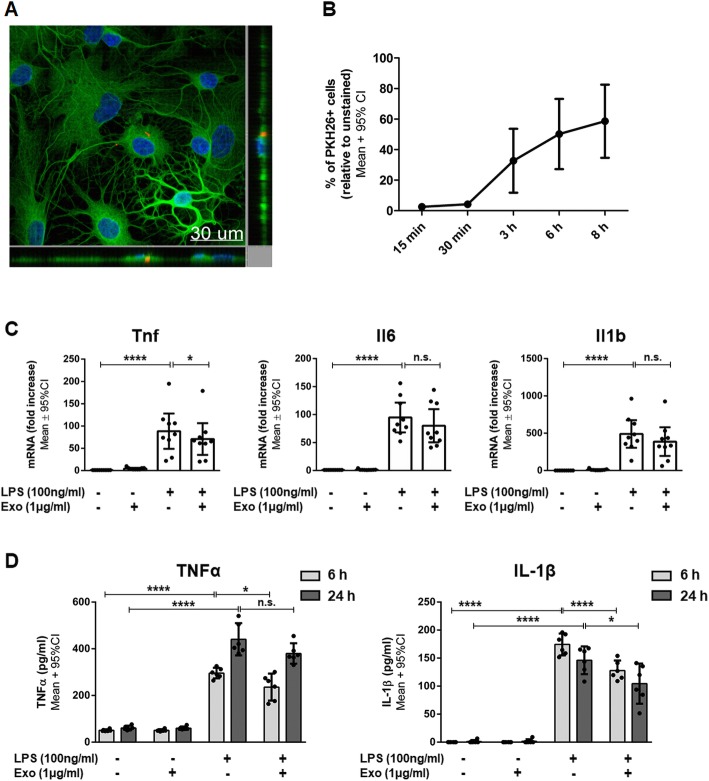
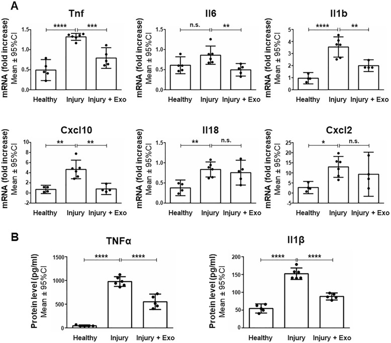
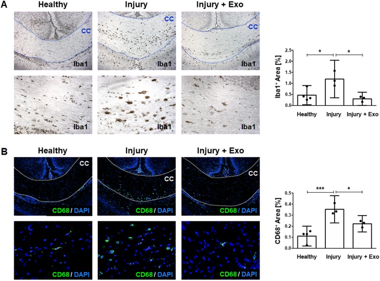
Background: Preterm newborns are at high risk of developing neurodevelopmental deficits caused by neuroinflammation leading to perinatal brain injury. Human Wharton's jelly mesenchymal stem cells (hWJ-MSC) derived from the umbilical cord have been suggested to reduce neuroinflammation, in part through the release of extracellular vesicle-like exosomes. Here, we studied whether exosomes derived from hWJ-MSC have anti-inflammatory effects on microglia-mediated neuroinflammation in perinatal brain injury.
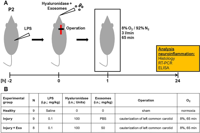
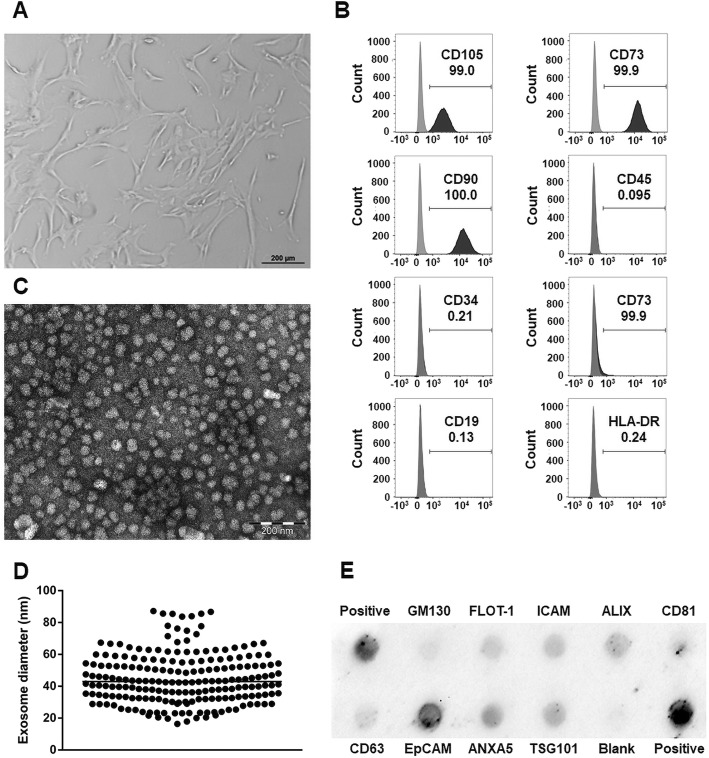
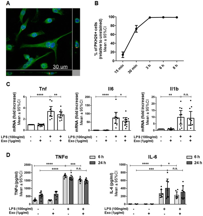
Methods: Using ultracentrifugation, we isolated exosomes from hWJ-MSC culture supernatants. In an in vitro model of neuroinflammation, we stimulated immortalized BV-2 microglia and primary mixed glial cells with lipopolysaccharide (LPS) in the presence or absence of exosomes. In vivo, we introduced brain damage in 3-day-old rat pups and treated them intranasally with hWJ-MSC-derived exosomes.
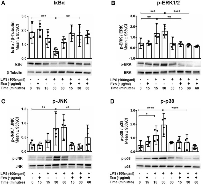
Results: hWJ-MSC-derived exosomes dampened the LPS-induced expression of inflammation-related genes by BV-2 microglia and primary mixed glial cells. The secretion of pro-inflammatory cytokines by LPS-stimulated primary mixed glial was inhibited by exosomes as well. Exosomes interfered within the Toll-like receptor 4 signaling of BV-2 microglia, as they prevented the degradation of the NFκB inhibitor IκBα and the phosphorylation of molecules of the mitogen-activated protein kinase family in response to LPS stimulation. Finally, intranasally administered exosomes reached the brain and reduced microglia-mediated neuroinflammation in rats with perinatal brain injury.
Conclusions: Our data suggest that the administration of hWJ-MSC-derived exosomes represents a promising therapy to prevent and treat perinatal brain injury.
Keywords: BV-2; Exosomes; Extracellular vesicles; Hypoxia-ischemia; Intranasal; Mesenchymal stem cells; Microglia; Neuroinflammation; Perinatal brain damage; Preterm birth; Umbilical cord; White matter injury.
Conflict of interest statement
Ethics approval and consent to participate
The study was approved by the Ethics Committee of the Canton of Bern (reference numbers: KEK BE 090_07 and KEK BE 178_03) and all patients involved gave written informed consent. All animal procedures were approved by the Veterinary Department of the Canton of Bern, Switzerland (reference number: BE117/16; 28'384).
Consent for publication
Not applicable.
Competing interests
The authors declare that they have no competing interests.
Publisher’s Note
Springer Nature remains neutral with regard to jurisdictional claims in published maps and institutional affiliations.
Figures
Similar articles
-
Intranasally Administered Exosomes from Umbilical Cord Stem Cells Have Preventive Neuroprotective Effects and Contribute to Functional Recovery after Perinatal Brain Injury.Cells. 2019 Aug 8;8(8):855. doi: 10.3390/cells8080855. Cells. 2019. PMID: 31398924 Free PMC article.
-
Intranasal Delivery of Umbilical Cord-Derived Mesenchymal Stem Cells Preserves Myelination in Perinatal Brain Damage.Stem Cells Dev. 2016 Aug 15;25(16):1234-42. doi: 10.1089/scd.2016.0027. Stem Cells Dev. 2016. PMID: 27392671
-
Human umbilical cord mesenchymal stem cell-derived exosomes attenuate neuroinflammation and oxidative stress through the NRF2/NF-κB/NLRP3 pathway.CNS Neurosci Ther. 2024 Mar;30(3):e14454. doi: 10.1111/cns.14454. Epub 2023 Sep 12. CNS Neurosci Ther. 2024. PMID: 37697971 Free PMC article.
-
Targeting Neuroinflammation in Preterm White Matter Injury: Therapeutic Potential of Mesenchymal Stem Cell-Derived Exosomes.Cell Mol Neurobiol. 2025 Mar 12;45(1):23. doi: 10.1007/s10571-025-01540-6. Cell Mol Neurobiol. 2025. PMID: 40072734 Free PMC article. Review.
-
Mesenchymal stem cell-derived exosomes regulate microglia phenotypes: a promising treatment for acute central nervous system injury.Neural Regen Res. 2023 Aug;18(8):1657-1665. doi: 10.4103/1673-5374.363819. Neural Regen Res. 2023. PMID: 36751776 Free PMC article. Review.
Cited by
-
Anti-inflammatory effects of antenatal administration of stem cell derived extracellular vesicles in the brain of rat fetuses with congenital diaphragmatic hernia.Pediatr Surg Int. 2023 Nov 13;39(1):291. doi: 10.1007/s00383-023-05578-9. Pediatr Surg Int. 2023. PMID: 37955723
-
Different Sourced Extracellular Vesicles and Their Potential Applications in Clinical Treatments.Cells. 2022 Jun 21;11(13):1989. doi: 10.3390/cells11131989. Cells. 2022. PMID: 35805074 Free PMC article. Review.
-
Pro-Inflammatory Priming of Umbilical Cord Mesenchymal Stromal Cells Alters the Protein Cargo of Their Extracellular Vesicles.Cells. 2020 Mar 16;9(3):726. doi: 10.3390/cells9030726. Cells. 2020. PMID: 32188006 Free PMC article.
-
Immunoregulatory Effects of Stem Cell-Derived Extracellular Vesicles on Immune Cells.Front Immunol. 2020 Feb 11;11:13. doi: 10.3389/fimmu.2020.00013. eCollection 2020. Front Immunol. 2020. PMID: 32117221 Free PMC article. Review.
-
Stem cell-based therapy as a promising approach in Alzheimer's disease: current perspectives on novel treatment.Cell Tissue Bank. 2021 Sep;22(3):339-353. doi: 10.1007/s10561-020-09896-3. Epub 2021 Jan 4. Cell Tissue Bank. 2021. PMID: 33398492 Review.
References
-
- Bajwa NM, Berner M, Worley S, Pfister RE. Population based age stratified morbidities of premature infants in Switzerland. Swiss Med Wkly. 2011;141:w13212. - PubMed
Publication types
MeSH terms
Substances
LinkOut - more resources
Full Text Sources
Other Literature Sources
