Review
. 2019 Mar 26;73(11):1317-1335.
doi: 10.1016/j.jacc.2018.12.054.

Artificial Intelligence in Cardiovascular Imaging: JACC State-of-the-Art Review

Affiliations
Review

Artificial Intelligence in Cardiovascular Imaging: JACC State-of-the-Art Review

Damini Dey et al. J Am Coll Cardiol. .

Abstract

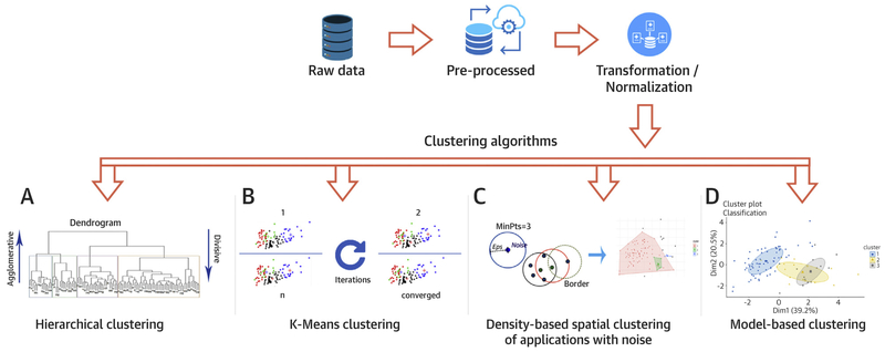
Data science is likely to lead to major changes in cardiovascular imaging. Problems with timing, efficiency, and missed diagnoses occur at all stages of the imaging chain. The application of artificial intelligence (AI) is dependent on robust data; the application of appropriate computational approaches and tools; and validation of its clinical application to image segmentation, automated measurements, and eventually, automated diagnosis. AI may reduce cost and improve value at the stages of image acquisition, interpretation, and decision-making. Moreover, the precision now possible with cardiovascular imaging, combined with "big data" from the electronic health record and pathology, is likely to better characterize disease and personalize therapy. This review summarizes recent promising applications of AI in cardiology and cardiac imaging, which potentially add value to patient care.

Keywords: artificial intelligence; cardiovascular imaging; deep learning; machine learning.


Figures

FIGURE 1
FIGURE 1. Steps in Performing Clustering Algorithm on the Data
The raw data is initially pre-processed and transformed, if needed. (A) Dendrogram of hierarchical clustering where height is the distance and each leaf represent a patient. The colored boxes represent the patients within the cluster. The number of clusters depends on where the dendrogram is cut. Agglomerative clustering is the bottom-up approach, where the patients are grouped in the higher hierarchy. Divisive clustering is the top-down approach where a single cluster is divided as it moves down the hierarchy. (B) Panel showing k-means cluster at multiple iterations. The algorithm calculates the centroid at each iteration and modulates the cluster until it converges. (C) Eps is the minimum distance between 2 points and MinPts is the minimum number of points to form a dense region. The algorithm searches the points based on these parameters and creates the cluster if the criteria are met. (D) Cluster analysis using model-based algorithm which detects 3 clusters in the mixture using Gaussian probabilistic model.
FIGURE 2
FIGURE 2. Topological Data Analysis for Isolating Patient Clusters
An existing published dataset containing clinical features of hospitalized patients with inpatient echocardiography utilization was used to create a network where clinically similar hospitalized subjects clustered in nodes and connected with overlapping subjects to form the edges. The topological network allows rapid visualization and interpretation of outcomes of interest. The distribution and frequency of echocardiograms performed in hospitalized patient clusters is shown in shades of red (A). The nodes are subsequently color coded with outcomes of interest like the length of stay (in days) to reflect visually any relationships with performance of echo-cardiography (B). The upper left region of the map (circle) shows an area where there is high utilization of echocardiography. Interestingly, this region has patients who are sicker, have longer length of stay (B), and also higher mortality (C).
FIGURE 3
FIGURE 3. Organization and Content of the REFINE SPECT Registry for the Purposes of Machine Learning (Blue)
Clinical data collection and analysis; (orange) imaging data collection and analysis; and (gray) integration of clinical and imaging databases. Reproduced with permission from Slomka et al. (71). MACE = major adverse cardiovascular events; MPI = myocardial perfusion imaging; QC = quality control; QGS = quantitative gated SPECT; QPS = quantitative perfusion SPECT; SPECT = single photon emission computed tomography.
FIGURE 4
FIGURE 4. ML to Predict All-Cause Mortality
Receiver-operating characteristic curves for prediction of death with 5-year follow-up compared to the Framingham risk score (FRS) and computed tomography angiography (CTA) severity scores (Segment Stenosis Score [SSS], Segment Involvement Score [SIS], modified Duke Index [DI]). *ML had significantly higher AUC than all other scores (P <0.001). Reproduced with permission from Motwani et al. (23). AUC = area under the curve; ML = machine learning.
FIGURE 5
FIGURE 5. Prediction of Lesion-Specific Ischemia by the Integrated Ischemia Risk Score by ML-Combined
(A) ML-combined versus quantitative plaque volumes (LD-NCP [low density noncalcified plaque], NCP, and total plaque volume). (B) ML-combined versus quantitative stenosis and pre-test likelihood of coronary artery disease. ML-combined had a significantly higher AUC compared with individual quantitative CTA plaque measures or the pre-test likelihood. *indicates AUC significantly different (p < 0.05) than that from the other measures. Reproduced with permission from Lee et al. (12). Abbreviations as in Figure 4.
FIGURE 6
FIGURE 6. Information Gain for Age, Sex, and Quantitative CTA Measures for Lesion-Specific Ischemia
In the left panel, measures directly related to plaque volumes are in light blue and the remaining measures are in dark blue. Variables with information gain >0.001 were used in machine learning. Contrast density difference had the highest information gain among quantitative CTA metrics. Reproduced with permission from Dey et al. (75). The right panel shows an example of the machine learning prediction of lesion-specific ischemia in a patient undergoing CTA. NCP and CP are shown in red and yellow image overlay in the left anterior descending (LAD) artery of a 67-year-old male symptomatic patient undergoing CTA, along with the integrated machine learning ischemia risk score (60% in the LAD). Invasive FFR measured in the LAD artery was 0.73. FFR = fractional flow reserve; LCX = left circumflex artery; RCA = right coronary artery; other abbreviations as in Figures 4 and 5.
FIGURE 7
FIGURE 7. Training of a Deep Convolutional Neural Network
Patterns of SPECT perfusion defects are identified by feature extraction (left) into a deep learning process (center) that combines parameters of location, shape, and density. This generates a probability of obstructive coronary artery disease in the left anterior descending artery (LAD), left circumflex artery (LCx), and right coronary artery (RCA) territories (right), which is trained by obstructive stenosis correlations by invasive coronary angiography. FC = fully connected layer; Max-pooling = filter that retains only the maximum value in a 2 × 2 patch; QPS = quantitative perfusion SPECT; ReLU = rectified linear unit (linear function mapping input to output values with a threshold). Adapted with permission from Betancur et al. (82).
FIGURE 8
FIGURE 8. The ROC Curves Comparing the ML Algorithm Versus Ischemic TPD and Expert Visual SDS for Predicting Revascularization
Reproduced with permission from Arsanjani et al. (88). ML = machine learning; ROC = receiver-operating curve; TPD = total perfusion deficit; VSDS = visual summed difference score.
FIGURE 9
FIGURE 9. Observed Proportion of Events and Predicted ML Score Grouped by Every Fifth Percentile of Risk Blue bars
indicate observed proportion of events, and orange points indicate predicted ML. Adapted with permission from Betancur et al. (70). MACE = major adverse cardiovascular events; ML = machine learning.

References

    1. Russell S, Norvig P. Artificial Intelligence: A Modern Approach. 2nd edition Upper Saddle River, New Jersey: Prentice Hall, 2003.
    1. Szolovits P, Patil RS, Schwartz WB. Artificial intelligence in medical diagnosis. Ann Intern Med 1988;108:80–7. - PubMed
    1. Darcy AM, Louie AK, Roberts LW. Machine learning and the profession of medicine. JAMA 2016;315:551–2. - PubMed
    1. Nilsson N The Quest for Artificial Intelligence: A History of Ideas and Achievements. New York: Cambridge University Press, 2009.
    1. Obermeyer Z, Emanuel EJ. Predicting the future - big data, machine learning, and clinical medicine. N Engl J Med 2016;375:1216–9. - PMC - PubMed

Publication types