

Age-Normative Pathways of Striatal Connectivity Related to Clinical Symptoms in the General Population
- PMID: 30898336
- PMCID: PMC6534442
- DOI: 10.1016/j.biopsych.2019.01.024
Age-Normative Pathways of Striatal Connectivity Related to Clinical Symptoms in the General Population
Abstract
Background: Altered striatal development contributes to core deficits in motor and inhibitory control, impulsivity, and inattention associated with attention-deficit/hyperactivity disorder and may likewise play a role in deficient reward processing and emotion regulation in psychosis and depression. The maturation of striatal connectivity has not been well characterized, particularly as it relates to clinical symptomatology.
Methods: Resting-state functional connectivity with striatal subdivisions was examined for 926 participants (8-22 years of age, 44% male) from the general population who had participated in two large cross-sectional studies. Developing circuits were identified and growth charting of age-related connections was performed to obtain individual scores reflecting relative neurodevelopmental attainment. Associations of clinical symptom scales (attention-deficit/hyperactivity disorder, psychosis, depression, and general psychopathology) with the resulting striatal connectivity age-deviation scores were then tested using elastic net regression.
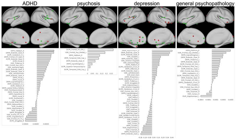
Results: Linear and nonlinear developmental patterns occurred across 231 striatal age-related connections. Both unique and overlapping striatal age-related connections were associated with the four symptom domains. Attention-deficit/hyperactivity disorder severity was related to age-advanced connectivity across several insula subregions, but to age-delayed connectivity with the nearby inferior frontal gyrus. Psychosis was associated with advanced connectivity with the medial prefrontal cortex and superior temporal gyrus, while aberrant limbic connectivity predicted depression. The dorsal posterior insula, a region involved in pain processing, emerged as a strong contributor to general psychopathology as well as to each individual symptom domain.
Conclusions: Developmental striatal pathophysiology in the general population is consistent with dysfunctional circuitry commonly found in clinical populations. Atypical age-normative connectivity may thereby reflect aberrant neurodevelopmental processes that contribute to clinical risk.
Keywords: ADHD; Depression; Development; General psychopathology; Psychosis; Striatum.
Copyright © 2019 Society of Biological Psychiatry. Published by Elsevier Inc. All rights reserved.
Conflict of interest statement
Disclosures
Dr. Malhotra has served as a consultant for Forum Pharmaceuticals and has served on a scientific advisory board for Genomind, Inc. Dr. Lencz has served as a consultant to Genomind, Inc. The other authors report no biomedical financial interests or potential conflicts of interest.
Figures


Similar articles
-
Corticostriatal causality analysis in children and adolescents with attention-deficit/hyperactivity disorder.Psychiatry Clin Neurosci. 2024 May;78(5):291-299. doi: 10.1111/pcn.13650. Epub 2024 Mar 5. Psychiatry Clin Neurosci. 2024. PMID: 38444215 Free PMC article.
-
Impaired functional connectivity within and between frontostriatal circuits and its association with compulsive drug use and trait impulsivity in cocaine addiction.JAMA Psychiatry. 2015 Jun;72(6):584-92. doi: 10.1001/jamapsychiatry.2015.1. JAMA Psychiatry. 2015. PMID: 25853901
-
Differential Resting-State Functional Connectivity of Striatal Subregions in Bipolar Depression and Hypomania.Brain Connect. 2016 Apr;6(3):255-65. doi: 10.1089/brain.2015.0396. Epub 2016 Jan 29. Brain Connect. 2016. PMID: 26824737 Free PMC article.
-
Atypical prefrontal connectivity in attention-deficit/hyperactivity disorder: pathway to disease or pathological end point?Biol Psychiatry. 2011 Jun 15;69(12):1168-77. doi: 10.1016/j.biopsych.2011.03.022. Epub 2011 May 5. Biol Psychiatry. 2011. PMID: 21546000 Review.
-
Differentiating frontostriatal and fronto-cerebellar circuits in attention-deficit/hyperactivity disorder.Biol Psychiatry. 2011 Jun 15;69(12):1178-84. doi: 10.1016/j.biopsych.2010.07.037. Epub 2010 Oct 20. Biol Psychiatry. 2011. PMID: 20965496 Review.
Cited by
-
Alterations of spontaneous brain activity in type 2 diabetes mellitus without mild cognitive impairment: a resting-state functional magnetic resonance study.Front Hum Neurosci. 2024 Jan 11;17:1305571. doi: 10.3389/fnhum.2023.1305571. eCollection 2023. Front Hum Neurosci. 2024. PMID: 38273877 Free PMC article.
-
Increased Reward-Related Activation in the Ventral Striatum During Stress Exposure Associated With Positive Affect in the Daily Life of Young Adults With a Family History of Depression. Preliminary Findings.Front Psychiatry. 2021 Jan 18;11:563475. doi: 10.3389/fpsyt.2020.563475. eCollection 2020. Front Psychiatry. 2021. PMID: 33584359 Free PMC article.
-
Associations between peripheral inflammation and resting state functional connectivity in adolescents.Brain Behav Immun. 2021 Jul;95:96-105. doi: 10.1016/j.bbi.2021.02.018. Epub 2021 Feb 23. Brain Behav Immun. 2021. PMID: 33631285 Free PMC article.
-
Is genetic risk of ADHD mediated via dopaminergic mechanism? A study of functional connectivity in ADHD and pharmacologically challenged healthy volunteers with a genetic risk profile.Transl Psychiatry. 2022 Jun 29;12(1):264. doi: 10.1038/s41398-022-02003-y. Transl Psychiatry. 2022. PMID: 35768414 Free PMC article.
-
Modulation of striatal functional connectivity differences in adults with and without autism spectrum disorder in a single-dose randomized trial of cannabidivarin.Mol Autism. 2021 Jul 1;12(1):49. doi: 10.1186/s13229-021-00454-6. Mol Autism. 2021. PMID: 34210360 Free PMC article. Clinical Trial.
References
-
- Di Martino A, Scheres A, Margulies DS, Kelly AM, Uddin LQ, Shehzad Z, et al. (2008): Functional connectivity of human striatum: a resting state FMRI study. Cerebral cortex. 18:2735–2747. - PubMed
-
- Postuma RB, Dagher A (2006): Basal ganglia functional connectivity based on a meta-analysis of 126 positron emission tomography and functional magnetic resonance imaging publications. Cerebral cortex. 16:1508–1521. - PubMed
-
- Haber SN (2003): The primate basal ganglia: parallel and integrative networks. Journal of chemical neuroanatomy. 26:317–330. - PubMed
-
- Alexander GE, DeLong MR, Strick PL (1986): Parallel organization of functionally segregated circuits linking basal ganglia and cortex. Annual review of neuroscience. 9:357–381. - PubMed
