Review
. 2019 Jul:127:303-311.
doi: 10.1016/j.nbd.2019.03.015. Epub 2019 Mar 18.

Multiscale recordings reveal the dynamic spatial structure of human seizures

Affiliations
Review

Multiscale recordings reveal the dynamic spatial structure of human seizures

Catherine A Schevon et al. Neurobiol Dis. 2019 Jul.

Abstract

The cellular activity underlying human focal seizures, and its relationship to key signatures in the EEG recordings used for therapeutic purposes, has not been well characterized despite many years of investigation both in laboratory and clinical settings. The increasing use of microelectrodes in epilepsy surgery patients has made it possible to apply principles derived from laboratory research to the problem of mapping the spatiotemporal structure of human focal seizures, and characterizing the corresponding EEG signatures. In this review, we describe results from human microelectrode studies, discuss some data interpretation pitfalls, and explain the current understanding of the key mechanisms of ictogenesis and seizure spread.

Keywords: Epilepsy; Focal seizures; Human single unit activity; Seizure localization; Surround inhibition.


Conflict of interest statement

Competing Interests: The authors have no competing interests to declare.

Figures

Figure 1:
Figure 1:
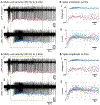
Ictal wavefront and tonic-clonic transition in a human seizure recorded with microelectrodes. This recording from a Utah microelectrode array implanted in the seizure onset zone of a patient with focal neocortical temporal lobe epilepsy demonstrates the progression of seizure invasion following global seizure onset, indicated by the initial pair of discharges. Data from four channels spaced ~800 microns apart are shown, filtered into low frequencies (1–50 Hz, black) and multiunit activity (0.5 – 3 kHz) with traces superimposed. The wavefront is clearly visible in MUA, but leaves little trace in the low frequencies and was entirely invisible to adjacent clinical electrodes. A slow expansion of the invaded territory can be seen. The tonic firing converts after 2–3 seconds to clonic bursting that synchronizes across the invaded territory.
Figure 2:
Figure 2:
Schematic descriptions of the dual-territory hypothesis. A) Relationship between the seizure focus (black disc), ictal core (blue), and penumbra (green). On standard visual EEG review, all three territories would appear as a single monolithic event. The seizure onset zone, determined typically from visualized EEG, may include both core and penumbra territories. As the seizure evolves, the penumbra expands faster than the core, and covers a much greater area. B) The transition between core and penumbra is the ictal wavefront, a brief period of intense, tonic firing with little, if any, manifestation in EEG. Excitatory barrages emanating from the wavefront into both core and penumbra territories result in dramatically different neuronal firing characteristics, depending on the status of inhibition. EEG, however, will show seizure-specific abnormalities in both territories, making it difficult to distinguish them clinically.
Figure 3:
Figure 3:
Action potential waveshape changes at seizure onset in a human hippocampal seizure. Microwire recordings from two Behnke-Fried depth arrays about 1 cm apart captured the ictal transition, each demonstrating a highly visible unit. A) Onset of tonic firing at different times, after which gradual reduction in amplitude of each unit is evident. B) Raw and z-scored voltage plots demonstrate a lag in the timing of amplitude reduction between the blue and red units, indicating that this is likely related to ictal activity rather than extracerebral factors.

Similar articles

Cited by

References

    1. Ahmed OJ, Kramer MA, Truccolo W, Naftulin JS, 2014. Inhibitory single neuron control of seizures and epileptic traveling waves in humans. BMC ….
    1. Aiba I, Noebels JL, 2015. Spreading depolarization in the brainstem mediates sudden cardiorespiratory arrest in mouse SUDEP models. Sci. Transl. Med 7, 282ra46 10.1126/scitranslmed.aaa4050 - DOI - PMC - PubMed
    1. Alarcon G, Garcia Seoane J, Binnie C, Martin Miguel M, Juler J, Polkey C, Elwes R, Ortiz Blasco J, 1997. Origin and propagation of interictal discharges in the acute electrocorticogram. Implications for pathophysiology and surgical treatment of temporal lobe epilepsy. Brain 120, 2259–2282. - PubMed
    1. Alfonsa H, Merricks EM, Codadu NK, Cunningham MO, Deisseroth K, Racca C, Trevelyan AJ, 2015. The contribution of raised intraneuronal chloride to epileptic network activity. J. Neurosci 35, 7715–7726. - PMC - PubMed
    1. Babb TL, Crandall PH, 1976. Epileptogenesis of human limbic neurons in psychomotor epileptics. Electroencephalogr. Clin. Neurophysiol 40, 225–243. - PubMed

Publication types