. 2019 Mar 22;363(6433):1314-1319.
doi: 10.1126/science.aav2914.

Autologous grafting of cryopreserved prepubertal rhesus testis produces sperm and offspring

Affiliations

Autologous grafting of cryopreserved prepubertal rhesus testis produces sperm and offspring

Adetunji P Fayomi et al. Science. .

Abstract

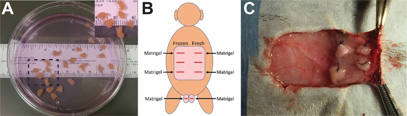
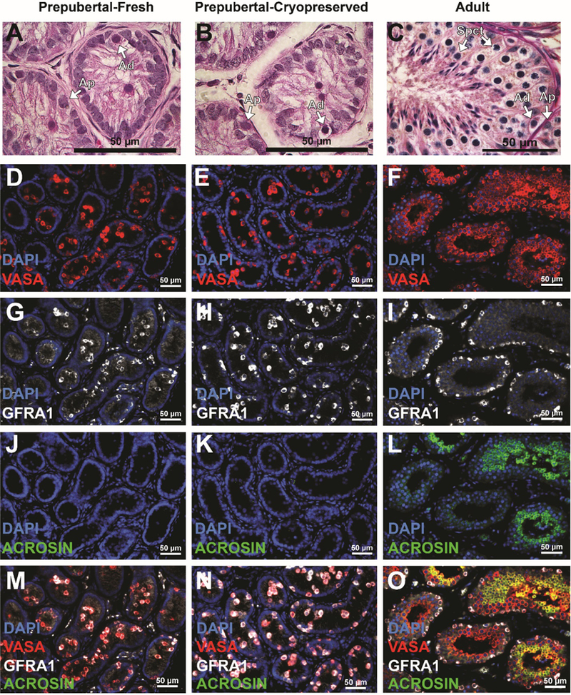
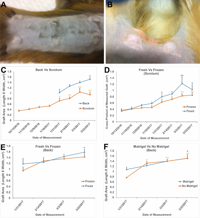
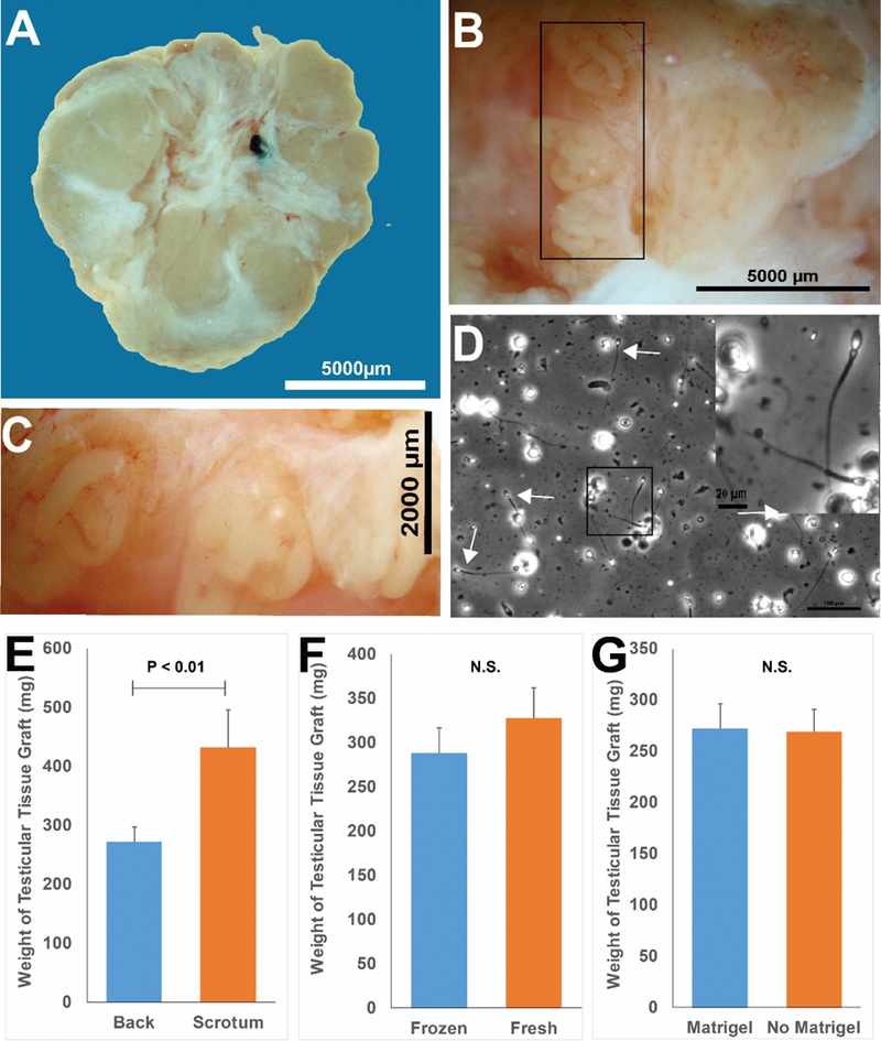
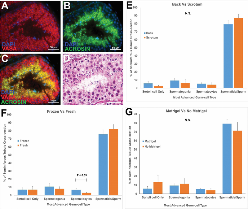
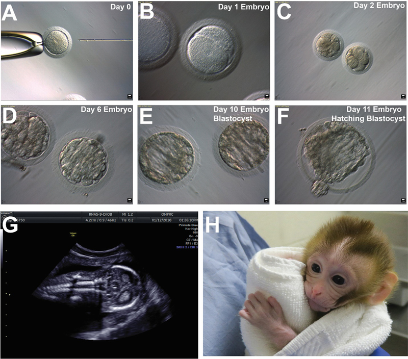
Testicular tissue cryopreservation is an experimental method to preserve the fertility of prepubertal patients before they initiate gonadotoxic therapies for cancer or other conditions. Here we provide the proof of principle that cryopreserved prepubertal testicular tissues can be autologously grafted under the back skin or scrotal skin of castrated pubertal rhesus macaques and matured to produce functional sperm. During the 8- to 12-month observation period, grafts grew and produced testosterone. Complete spermatogenesis was confirmed in all grafts at the time of recovery. Graft-derived sperm were competent to fertilize rhesus oocytes, leading to preimplantation embryo development, pregnancy, and the birth of a healthy female baby. Pending the demonstration that similar results are obtained in noncastrated recipients, testicular tissue grafting may be applied in the clinic.


Figures

Figure 1:
Figure 1:. Autologous grafting ofprepubertal testicular tissue fragments.
Fresh or cryopreserved testicular tissue fragments (9–20 mm3) from prepubertal monkeys (A). (A, Inset) Higher magnification image of area demarcated with dashed black box in (A). Testicular tissue fragments were grafted under the back skin or scrotal skin, as indicated in (B; viewed from the back of animal). Matrigel was added to four of the six graft sites on the back and both scrotal sites (B). Four pieces offresh or frozen-thawed testis tissue were sutured to the subcutaneous aspect of the skin at each graft site (C).
Figure 2:
Figure 2:. Histological and immunofluorescent analyses ofprepubertal testicular tissue prior to grafting.
Hematoxylin and Eosin staining indicate thatpre-graftedfresh (A) and frozen-thawed (B) testis tissues are immature. In contrast, multiple layers of germ cells with complete spermatogenesis were seen in adult testis tissue controls (C). Arrows indicate undifferentiated type Adark (Ad) and Apale (Ap) stem/progenitor spermatogonia and spermatocytes (Spct). Immunofluorescence staining for VASA+ germ cells (red; D-F); GFRA1 + undifferentiated spermatogonia (white; G-I) and ACROSIN+ post-meiotic germ cells (green; J-L). Merged images are shown in (M-O). Scale bar = 50μm. Please see additional staining for UTF1, BOULE and CREM in Supplementary Figure S2.
Figure 3:
Figure 3:. Testicular tissue grafts increase in size during the 8-to 12-month in vivo incubation period.
By 4–5 months after grafting, grafts were easily visualized under the back skin (A) and scrotal skin (B). Calipers were used to monitor graft growth (C-F). All grafts grew during the 8-to 12-month incubation period. Grafts on the back and in the scrotal area grew significantly over time relative to the first graft size measurement (C). Graft sizes (length x width) on each analysis date were not impacted by processing (fresh versus frozen-thawed) in the scrotum (D) or on the back (E) or by addition of Matrigel to the back sites (F). Frozen grafts on the back exhibited a trend toward increasing size through the experiment (E), but that increase was not statistically significant. All other grafts exhibited statistically significant increases in size through the experiment. Data points are presented as mean ± standard error of the mean. * indicates P<0.05 compared with initial graft size measurement and ** indicates P<0.01 compared with initial graft size measurement within each treatment group.
Figure 4:
Figure 4:. Recovery of testicular tissue grafts.
Grafts were recovered as one fused tissue, but white fibrous tissue may demarcate margins between individual testis tissue pieces that were originally placed at each graft (A). Seminiferous tubules could be distinguished after gentle teasing apart of the graft tissue with forceps (B). (C) Higher magnification of black box area in B. Panel D (white arrows) depicts sperm that were released by mechanical dissection of a scrotal graft from animal 13–030. (D, Inset) Higher magnification of black box region in (D). Grafts recovered from under the scrotal skin were larger than those recovered from under the back skin (E). Graft size was not impacted by processing (fresh versus frozen, (E)) or by addition of Matrigel (F). Bar graphs are presented as mean ± standard error of the mean. P<0.05 was considered by be significant. N.S. = not significant.
Figure 5:
Figure 5:. Histological evaluation of spermatogenic development in grafts.
Immunofluorescence staining of recovered grafted tissue for VASA+ germ cells (red, A) and ACROSIN+ post-meiotic cells (green, B). DAPI counterstain marks all cell nuclei (blue). The merged VASA/ACROSIN/DAPI co-stain is shown in (C). Please see Figure S4 for additional markers of undifferentiated spermatogonia (UTF1), spermatocytes (BOULE) and spermatids (CREM). Hematoxylin and Eosin staining of post-graft tissues (D). Please see H and E staining for grafts from each individual animal in Figure S5. Quantification of most advanced germ cell type in graft seminiferous tubules (E-G). Bars represent mean ± standard error of the mean. N.S. = not significant; P<0.05 was considered statistically significant.
Figure 6:
Figure 6:. Functional evaluation of graft-derived sperm.
Sperm were derived from a cryopreserved graft retrieved from the left scrotum of13–008 that was recovered nine months after grafting. Fresh graft-derived sperm were used for the May 2017ICSI trial. The remaining sperm were cryopreserved and used for the October 2017 and November 2017 trials (See Table S2). Graft-derived sperm were used to fertilize Rhesus oocytes by ICSI (A). The resulting embryos attained 2-cell stage by day 1 (B); 8-cell stage by day 2 (C); morula stage by day 6 (D); blastocyst stage by day 10 (E); and hatching blastocyst by day 11 in culture (F). Blastocyst embryos were transferred to recipient females and a pregnancy was confirmed by ultrasound on December 12, 2017. Normal fetal development was confirmed by ultrasound on January 15, 2018 (G) and a graft-derived baby (Grady) was born by c-section on April 16, 2018 (H, image from two-week check-up).

Comment in

References

    1. Green DM et al. , Fertility of male survivors of childhood cancer: a report from the Childhood Cancer Survivor Study. Journal of clinical oncology : official journal of the American Society of Clinical Oncology 28, 332–339 (2010). - PMC - PubMed
    1. Levine J, Canada A, Stern CJ, Fertility preservation in adolescents and young adults with cancer. Journal of clinical oncology : official journal of the American Society of Clinical Oncology 28, 4831–4841 (2010). - PubMed
    1. Meistrich ML, Male gonadal toxicity. Pediatric Blood & Cancer 53, 261–266 (2009). - PMC - PubMed
    1. Wallace WH, Anderson RA, Irvine DS, Fertility preservation for young patients with cancer: who is at risk and what can be offered? The lancet oncology 6, 209–218 (2005). - PubMed
    1. Goossens E, Van Saen D, Tournaye H, Spermatogonial stem cell preservation and transplantation: from research to clinic. Human Reproduction 28, 897–907 (2013). - PubMed

Publication types