. 2019 Mar 21;9(1):4973.
doi: 10.1038/s41598-019-41426-4.

A complex of novel protease inhibitor, ovostatin homolog, with its cognate proteases in immature mice uterine luminal fluid

Affiliations

A complex of novel protease inhibitor, ovostatin homolog, with its cognate proteases in immature mice uterine luminal fluid

Hsien-Lu Huang et al. Sci Rep. .

Abstract

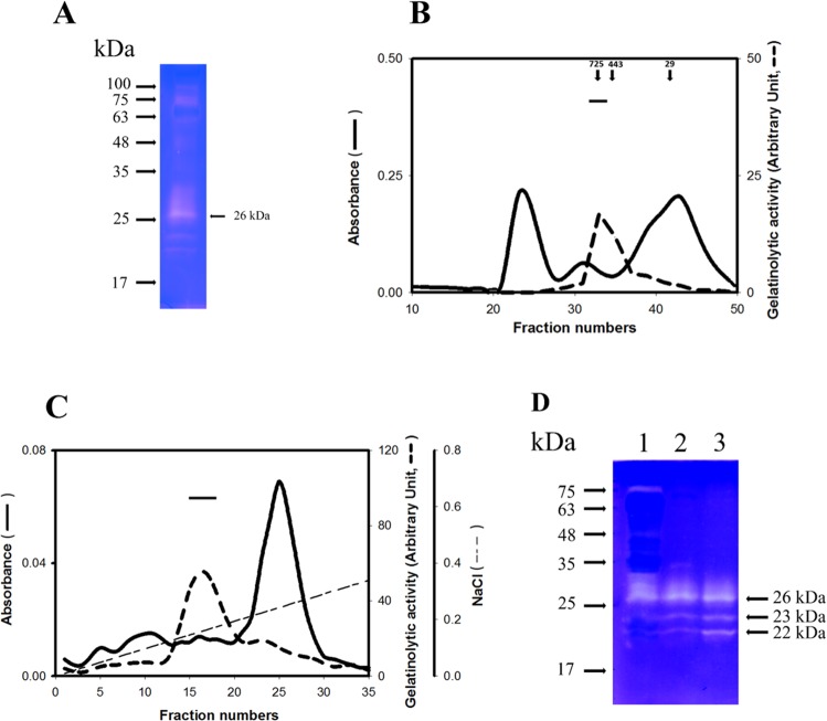
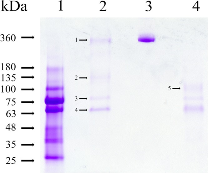
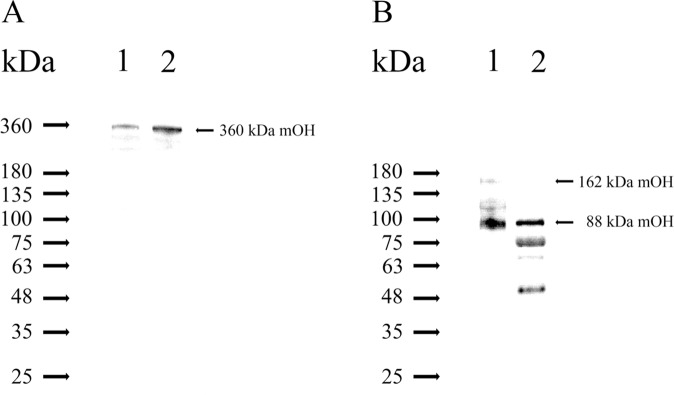
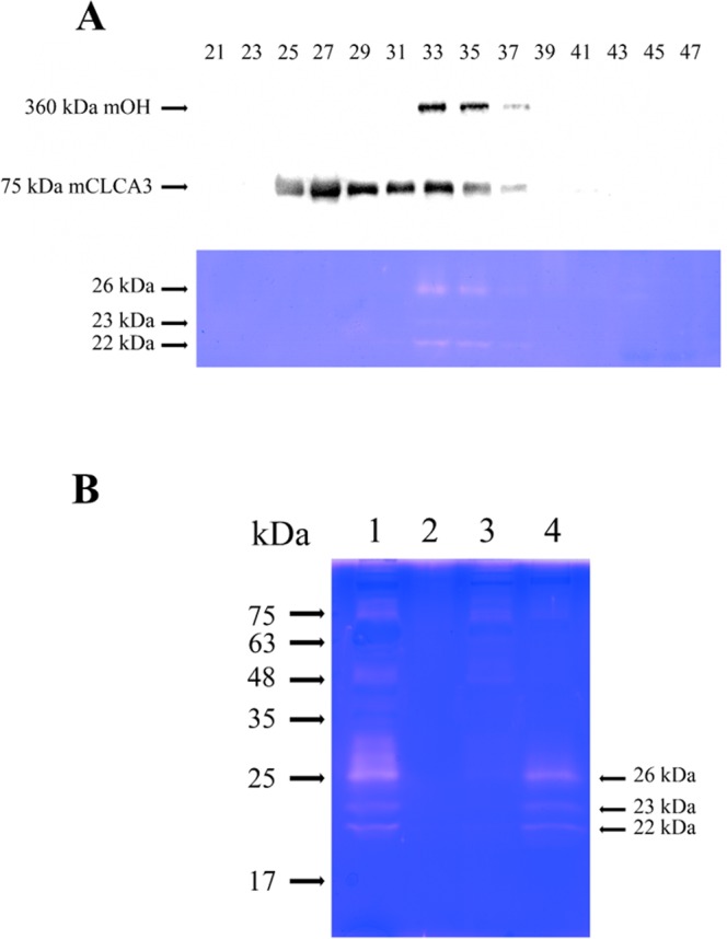
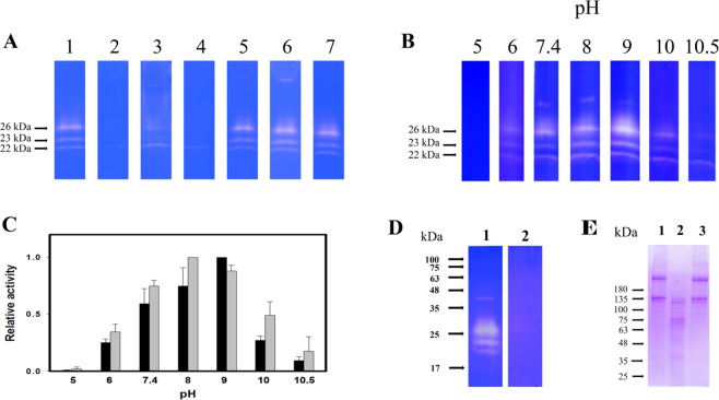
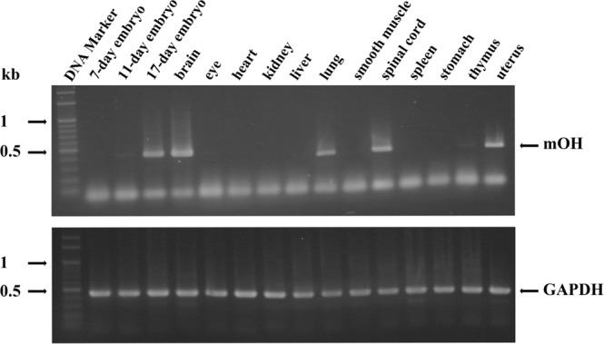
A predominant gelatinolytic enzyme with approximately 26 kDa was observed in gelatin zymogram of immature mice uterine luminal fluid (ULF). Size exclusion analysis revealed that the native size of this enzyme was close to that of human α2-macroglobulin (α2-MG), a 725 kDa protein. This large protease was isolated by a series of chromatographic steps on the Sephacryl S-400 and DEAE-Sepharose columns. The results from gelatin zymography and SDS-PAGE analysis supported that this large protease consists of gelatinolytic enzyme and a 360 kDa protein. Through tandem mass spectrometry analysis followed by MASCOT database search, the 360 kDa protein was identified as ovostatin homolog (accession: NP_001001179.2) assigned as a homolog of chicken ovostatin, a protease inhibitor. The co-fractionation analysis by gel filtration and mouse ovostatin homolog (mOH) co-immunoprecipitation experiments demonstrated that the mOH formed a complex with three gelatinolytic enzymes in immature mice ULF. Substrate zymography analysis revealed that the mOH-associated gelatinolytic enzymes were suitable to digest type I collagen rather than type IV collagen. In addition, the refolded mOH-associated 26 kDa gelatinolytic enzyme displayed the type I collagen-digesting activity in the assay, but the other two enzymes did not have this function. RT-PCR analysis showed that mOH gene was abundantly expressed in brain, spinal cord, lung, uterus, and in 17-day embryo. Taken together, our data suggest that mOH/cognate protease system may play a potential role in regulation of tissue remodeling and fetal development.


Conflict of interest statement

The authors declare no competing interests.

Figures

Figure 1
Figure 1
Purification of the ULF gelatinolytic enzyme. (A) Gelatin zymogram of immature mouse ULF. The ULF (6 μl) was analyzed by gelatin zymography. (B) Elution profile of ULF gelatinolytic enzyme carried out by gel filtration chromatography. Soluble ULF (1 ml) was subjected to gel filtration chromatography on a Sephacryl S-400 column. The elution was monitored by UV spectrophotometry with absorbance at 280 nm (—). The gelatinolytic activity of 26 kDa enzyme in each indicated fraction was quantitatively detected (---). Arrows indicate the protein markers eluted from the same column. The protein markers are human α2MG (725 kDa), apoferritin (443 kDa) and carbonic anhydrase (29 kDa). The horizontal bar represents the pooled fractions for further purification by the DEAE-Sepharose chromatography. (C) Elution profile of DEAE-Sepharose chromatography. The pooled fractions are marked by a horizontal bar. (D) Gelatin zymographic assay. Lane 1: immature mouse ULF (6 μl). Lane 2: the pooled fractions (0.3 ml) from gel filtration. Line 3: the pooled fractions (0.25 ml) from DEAE-Sepharose chromatography.
Figure 2
Figure 2
SDS-PAGE analysis of the gelatinolytic enzyme complex isolated from immature mice ULF. An aliquot was separated by a 4–15% gradient acrylamide gel with 0.1% SDS. Lane 1: ULF (5 μl) treated with 2-mercaptoethanol. Lane 2: the gelatinolytic enzyme complex (3 μg) without 2-mercaptoethanol treatment. Lane 3: human α2MG dimer (360 kDa, 3 μg). Lane 4: the gelatinolytic enzyme complex (3 μg) with 2-mercaptoethanol treatment. After electrophoresis, the gel was stained with Coomassie Brilliant Blue and then destained by 10% acetic acid. Five bands including 360, 135, 88, 75 and 66 kDa were numbered and indicated by the arrow bar.
Figure 3
Figure 3
mOH is present in immature mouse ULF. (A) The immature mouse ULF and the gelatinolytic enzyme complex were separated by non-reducing SDS-PAGE and then analyzed by Western blotting, probed for mOH by POHA. The POHA was purified from rabbit anti-mOH sera. Lane 1: immature mouse ULF (5 μl). Lane 2: the gelatinolytic enzyme complex (0.5 μg) that was the resulting product after immature mouse ULF was purified by gel filtration and DEAE-Sepharose chromatography. (B) The immature mouse ULF and the gelatinolytic enzyme complex were separated by reducing SDS-PAGE and then analyzed by Western blotting, probed for reducing mOH by rabbit anti-GST-mOH[870–940] sera. Lane 1: immature mouse ULF (5 μl). Lane 2: the gelatinolytic enzyme complex (0.5 μg).
Figure 4
Figure 4
A complex of mOH with three gelatinolytic enzymes in immature mouse ULF. (A) Co-elution of mOH with gelatinolytic enzymes during purification of mOH by gel filtration. Each indicated fraction (0.3 ml) was separated by SDS-PAGE and then applied to immunoblot analysis for detection of mOH protein using POHA (upper panel) and for detection of mCLCA3 using anti-mCLCA3 antibody (middle panel), respectively. The identical samples were also subjected to gelatin zymography for detection of gelatinolytic enzymes (lower panel). The original immunoblots and scan of gelatin zymogram were displayed in Supplementary Fig. S3. (B) Co-immunoprecipitation of mOH with three gelatinolytic enzymes. Either POHA or preimmune antibody (PIA) was pre-immobilized on Protein A-Sepharose (60 μl), followed by incubation with immature mice ULF (30 μl). After centrifugation, the pellet was re-suspended in 30 μl of sample buffer. An aliquot of re-suspended pellet or supernatant was subjected to SDS-PAGE, followed by gelatin zymography for detection of gelatinolytic enzymes. Lane 1: PIA-treated ULF supernatant (6 μl). Lane 2: PIA-treated ULF pellet (6 μl). Lane 3: POHA-treated ULF supernatant (6 μl), Lane 4: POHA-treated ULF pellet (6 μl).
Figure 5
Figure 5
Characterization of mOH-associated gelatinolytic enzyme activity. (A) The effects of protease inhibitors on the activity of mOH-associated gelatinolytic enzymes. An aliquot (0.3 ml) isolated from gel filtration was subjected to SDS-PAGE, followed by gelatin zymographic assay. After electrophoresis, the gel was sliced, and each sliced gel was incubated without protease inhibitor (lane 1) or with different protease inhibitors. The protease inhibitors were bezamidine (2 mM, lane 2); chymostatin, (100 μM, lane 3); leupeptin (100 μM, lane 4); pepstatin (20 μM, lane 5); iodoacetamide (0.1 mM, lane 6); 1-10-phenanthroline (1 mM, lane 7). The original scans of gelatin zymograms were shown in Supplementary Fig. S5. (B) The effects of pH titration on the activity of mOH-associated gelatinolytic enzymes. An aliquot (0.3 ml) isolated from gel filtration was separated by SDS-PAGE, followed by gelatin zymographic assay in the presence of different pH buffers, respectively. The buffers include 0.1 M sodium citrate (pH 5.0 and 6.0), 0.1 M Tris buffers (from pH 7.4 and 8.0) and 0.1 M glycine (pH 9, 10 and 10.5). The original scans of gelatin zymograms were shown in Supplementary Fig. S6. (C) Quantitation of relative gelatinolytic activities averaged from three independent experiments. The highest activity in pH titration represent 100%. The optimal pH for the activity of 26 kDa (black bar) and 22 kDa gelatinolytic enzymes (gray bar) is 9 and 8, respectively. Means ± SD, n = 3. (D) The substrate specificity of mOH-associated gelatinolytic enzymes. An aliquot (2 μg) of gelatinolytic enzymes complex from purification was carried out for gelatin zymography in the presence of different substrates, type I collagen (lane 1) and type IV collagen (lane 2), respectively. (E) The type I collagen degraded by mOH-associated 26 kDa gelatinolytic enzyme. The free 26 and 23/22 kDa enzymes were incubated with type I collagen, respectively, as described in “Materials and Methods”. After overnight incubation, an aliquot (8 μL) of supernatant from each reaction mixture was applied to analysis by SDS-PAGE. Lane 1: incubation with blank gel. Lane 2: incubation with mOH-free 26 kDa enzyme. Lane 3: incubation with mOH-free 23/22 kDa enzymes.
Figure 6
Figure 6
Tissue distribution of mOH mRNA. Total RNAs isolated from various adult mice tissues or from different stages of mice embryos were subjected to RT-PCR. The resulting products were separated in agarose gel and visualized by ethidium bromide staining under exposure of UV light. The RT-PCR product of GAPDH was served as the internal control. The original agarose gels were shown in Supplementary Fig. S7.

Similar articles

References

    1. Evans GS, Gibson DF, Roberts SA, Hind TM, Potten CS. Proliferative changes in the genital tissue of female mice during the oestrous cycle. Cell and Tissue Kinetics. 1990;23:619–635. - PubMed
    1. Rugh, R. Reproductive Systems of Adult Mice, New York: Oxford University Press. (1990).
    1. Heussen C, Dowdle EB. Electrophoretic analysis of plasminogen activators in polyacrylamide gels containing sodium dodecyl sulfate and copolymerized substrates. Anal Biochem. 1980;102:196–202. doi: 10.1016/0003-2697(80)90338-3. - DOI - PubMed
    1. Ricci S, D’Esposito V, Oriente F, Formisano P, Di Carlo A. Substrate-zymography: a still worthwhile method for gelatinases analysis in biological sample. Clin Chem Lab Med. 2016;54:1281–1290. - PubMed
    1. Rodgers WH, et al. Patterns of matrix metalloproteinase expression in cycling endometrium imply differential functions and regulation by steroid hormones. J Clin Invest. 1994;94:946–953. doi: 10.1172/JCI117461. - DOI - PMC - PubMed

Publication types