. 2019 Mar 21;9(1):4984.
doi: 10.1038/s41598-019-41478-6.

IL-27 promotes NK cell effector functions via Maf-Nrf2 pathway during influenza infection

Affiliations

IL-27 promotes NK cell effector functions via Maf-Nrf2 pathway during influenza infection

Pawan Kumar et al. Sci Rep. .

Abstract

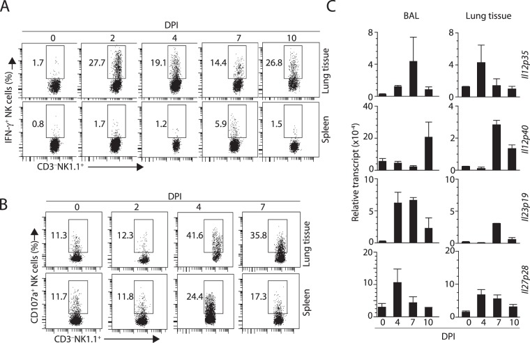
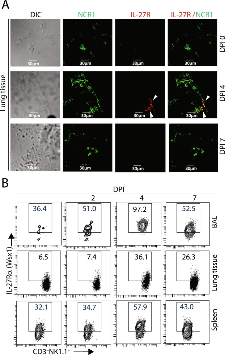
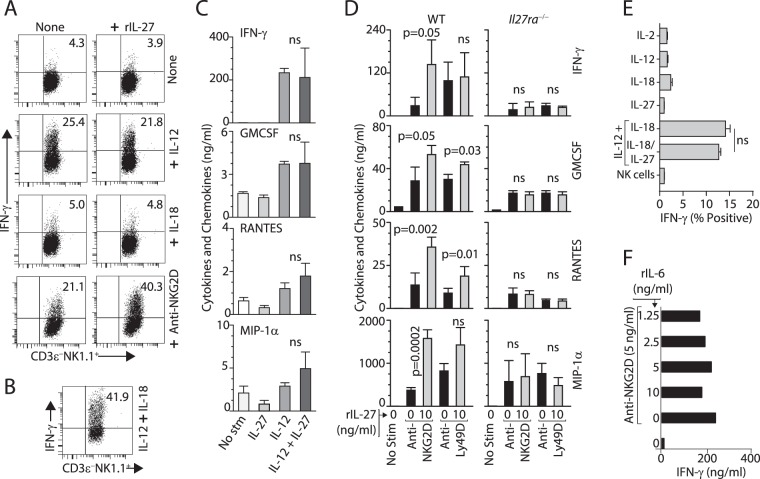
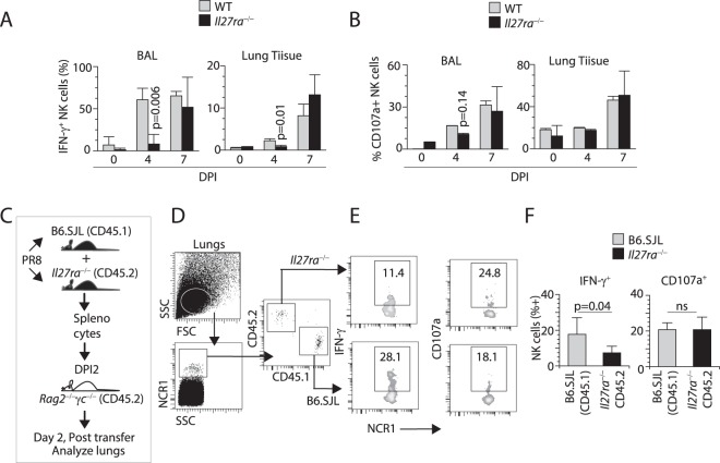
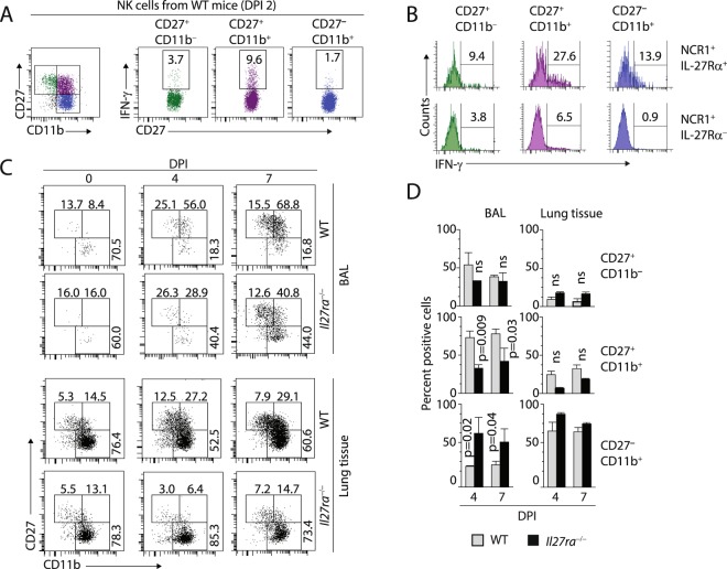
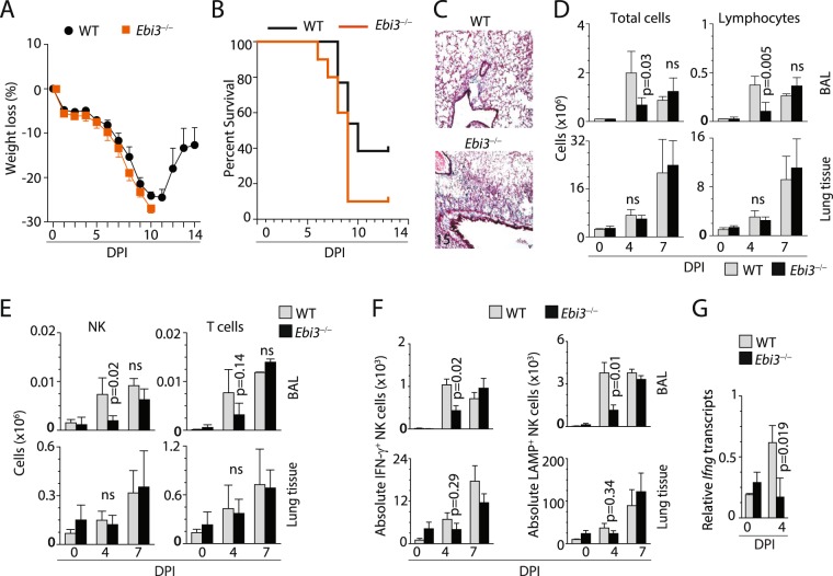
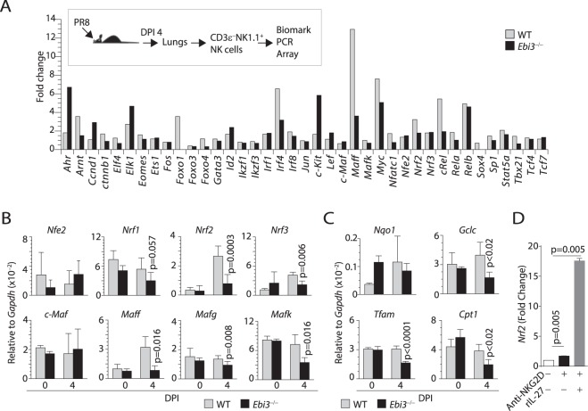
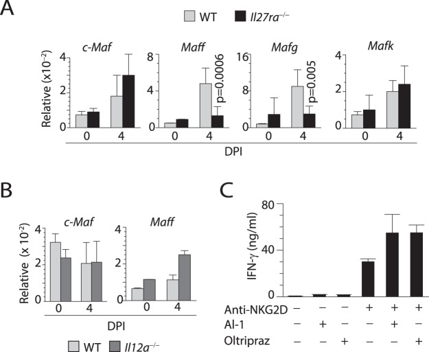
Influenza virus targets epithelial cells in the upper respiratory tract. Natural Killer (NK) cell-mediated early innate defense responses to influenza infection include the killing of infected epithelial cells and generation of anti-viral cytokines including interferon gamma (IFN-γ). To date, it is unclear how the underlying cytokine milieu during infection regulates NK cell effector functions. Our data show during influenza infection myeloid cell-derived IL-27 regulates the early-phase effector functions of NK cells in the bronchioalveolar and lung tissue. Lack of IL-27R (Il27ra-/-) or IL-27 (Ebi3-/-) resulted in impaired NK cell effector functions including the generation of anti-viral IFN-γ responses. We identify CD27+CD11b+ NK cells as the primary subset that expresses IL-27R, which predominantly produces IFN-γ within the upper respiratory tract of the infected mice. IL-27 alone was incapable of altering the effector functions of NK cells. However, IL-27 sensitizes NK cells to augment both in vitro and in vivo responses mediated via the NKG2D receptor. This 'priming' function of IL-27 is mediated partly via transcriptional pathways regulated by Mafs and Nrf2 transcriptionally regulating TFAM and CPT1. Our data for the first time establishes a novel role for IL-27 in regulating early-phase effector functions of NK cells during influenza infection.


Conflict of interest statement

The authors declare no competing interests.

Figures

Figure 1
Figure 1
NK cell infiltration and function coincides with IL-27p28 expression in the BAL and lung tissue of mice during the early phase influenza infection. (A) Production of IFN-γ peaks on DPI 4. Intracellular IFN-γ was analyzed in gated the lung tissue or spleens from the infected mice on indicated DPIs. (B) Expression of CD107a, a surrogate marker for the release of cytolytic granules including granzyme B peaks on DPI 4 in both lung tissue and spleens of the infected mice. (C) Expression of Il12p35, Il12p40, Il23p19, and Il27p28 transcripts in the BAL cells and lung tissue on different DPIs. Data shown are from two or three independent experiments with 4–7 mice (A), 3–4 mice (B) or 4 mice (C except for DPI7). DPI = Days post infection
Figure 2
Figure 2
Expression of IL-27R (Wsx-1) on NK cells during influenza infection. (A) Confocal analyses of IL-27R expression in NCR1+ NK cells in the lung tissue on indicated DPIs. Lung sections from infected mice were stained with anti-NCR1 and anti-Wsx-1 mAbs. (B) Flow cytometric analyses of IL-27R expression among CD3NK1.1+ NK cells obtained from BAL, lung tissue, or spleens of influenza-infected mice on different days of post-infection. Percent IL-27R (Wsx-1) positive cells are shown. Three mice each for A and B are analyzed, and representative images or data are presented.
Figure 3
Figure 3
IL-27 regulates NKG2D and Ly49D-mediated effector functions of NK cells. (A) IL-27 augments NKG2D- but not IL-12-mediated IFN-γ production. NK cells were cultured with IL-12 (10 ng/ml), IL-18 (10 ng/ml), or plate-bound anti-NKG2D mAb (5 ng/ml) for 12 hours and the production of IFN-γ was examined by intracellular staining. (B) IL-12 (1 ng/ml) in combination with IL-18 (10 ng/ml) stimulate NK cells to produce a significant amount of intracellular IFN-γ. (C) IL-27 regulates the production of multiple cytokines and chemokines. NK cells from WT mice were stimulated with IL-27 (10 ng/ml), IL-12 (10 ng/ml), or IL-27+ IL-12 (10 ng/ml each) for 18 hours and the supernatants were tested for the presence of indicated cytokines and chemokines. (D) Activation receptor-mediated inflammatory cytokine and chemokine production are regulated by IL-27. NK cells from WT or Il27ra−/− were stimulated with anti-NKG2D (5 ng/ml) or anti-Ly49D mAb (5 ng/ml) in the presence or absence of rIL-27 (10 ng/ml). (E) IL-27 does not mediate additive effect on IL-12 and IL-18-mediated IFN-γ production in NK cells. Purified NK cells were cultured with indicated individual or combinations of cytokines (10 ng/ml each) for 12 hours and the IFN-γ was examined by intracellular staining among CD3εNK1.1+ NK cells. (F) rIL-6 do not play a similar role as that of IL-27. Titrated concentrations rIL-6 (1.25 to 10 ng/ml) were used in the presence of mitogenic anti-NKG2D mAb (5 ng/ml). IL-2-cultured NK cells were used (AE). Data presented are a representative of 3–4 independent experiments generated from 3–4 mice per group per experiment.
Figure 4
Figure 4
IL-27 regulates NK cells effector function during influenza infection. (A) Lack of IL-27Rα significantly reduces the percentages of intracellular IFN-γ+ NK cells. Bar diagram of IFN-γ+ NK cells from BAL and lung tissues of WT and Il27ra−/− mice different DPI are shown. (B) Lack of IL-27Rα does not affect the cytotoxic potentials of NK cells as measured by its surrogate marker CD107a (Lamp1). NK cells from BAL and lung tissues of WT and Il27ra−/− mice different DPI are shown. (C) The schematic for the adoptive transfer of splenocytes from WT (CD45.1) and Il27ra−/− (CD45.2) mice into Rag2−/−γc−/− mice. Host Rag2−/−γc−/− mice were infected with influenza virus, and their splenocytes were analyzed on DPI 2. (D) Lungs from Rag2−/−γc−/− mice isolated and the CD3NCR1+ NK cells were analyzed. (E) Percent IFN-γ or CD107a (LAMP1)-positive NK cells from one representative mouse each are shown. (F) Average percent positive cells from four mice per genotype are shown. Data in A, B, D-F are averages with standard deviation and are obtained from a minimum of four individual mice and are representatives of at least two independent experiments.
Figure 5
Figure 5
Lack of IL-27Rα results in reduced number of CD27+CD11b+ NK cells subset during influenza infection. (A) CD27+CD11b+ NK cell subset predominantly produces IFN-γ during influenza infection. CD3NCR1+ NK cells from the lungs of influenza-infected WT mice on DPI 2 are sub-divided based on CD27 and CD11b (left), and the levels of IFN-γ+ was analyzed in each subset (right). (B) Among CD27+CD11b+ subset, IL-27Ra+ NK cells are the predominant producers of IFN-γ. NK cells from the lungs of WT mice on DPI 2 were analyzed based on CD27, CD11b, and IL-27Rα staining. (C) The number of CD27+CD11b+ NK cells are significantly reduced in the lungs of Il27ra−/− mice during influenza infection. CD3NCR1+ NK cells from BAL and lungs of WT and Il27ra−/− mice on indicated DPI were stained for the expression of CD27 and CD11b. (D) Bar diagram shows the percentage of different effector NK subsets in the BAL and lungs of WT and Il27ra−/− mice on DPI 4 and DPI 7. Data presented in A and B was a representative one mouse, which is a representative of 4–6 mice from two independent experiments. Data presented in D were an average of three mice per genotype.
Figure 6
Figure 6
Mice lacking EBI3 exhibit significantly impaired NK cell-mediated effector functions. (A) Mice that lack EBI3 do not recover their weight loss and succumb to influenza infection. WT and Ebi3−/− mice were infected with influenza virus, and their weight loss or (B) survival was monitored over the indicated DPIs. (C) Lack of EBI3 leads to increased inflammation as characterized by collagen deposition in the lung tissue (Mason’s trichrome staining, blue). (D) Lack of EBI3 decreases the total number of lymphocytes recruited to the lung. (E) Total number of NK cells recruited to the lungs were decreased in mice lacking EBI3. (F) An absolute number of NK cells producing IFN-γ or expressing CD107a (LAMP1) were significantly decreased in the absence of EBI3. (G) The reduction in the production of IFN-γ is due to an impaired transcription of Ifng gene. NK cells were isolated on indicated DPIs from the lungs of WT and Ebi3−/− mice, total RNA was isolated, and used to quantify the abundance of the transcripts. Data were generated from two independent experiments with 8–10 (A), 3–6 (B), 3–5 (C,D) or 3–4 (E, except DPI 0) mice per group.
Figure 7
Figure 7
IL-27 regulates MAFF and Nrf2 expression during influenza infection. (A) NK cells from the influenza-infected WT and Ebi3−/− mice on DPI 4 were analyzed for the expression levels of a panel of transcription factors using Fluidigm Transcription Factor arrays. (B) RT-qPCR data shows expression of Nfe2, Nrf1, Nrf2, Nrf3, cMaf, Maff, Mafg, and Mafk on sorted NK cells from WT and Ebi3−/− mice on day four post influenza infection. (C) RT-qPCR data showing reduced expression of Nqo1, Ho, Tfam1, and Cpt1 on sorted NK cells from WT and Ebi3−/− mice on day four post influenza infection. (D) NK cells were stimulated with plate-bound anti-NKG2D mAb in the presence or absence of rIL-27 and the mRNA were analyzed for the expression levels of Nrf2. Gene array data were generated using Fluidigm 50-selected gene array from sorted NK cells of WT (n = 2) and Ebi3−/− (n = 2) day four influenza infected or control mock-infected mice. Data in (B) and (C) generated from 4 mice per genotype (except mock-infected mice).
Figure 8
Figure 8
Lack of IL-27Ra but not IL-12Ra results in reduced MAF activation during influenza infection. (A) RT-qPCR data shows expression of cMaf, Maff, Mafg, and Mafk in sorted NK cells from WT and Il27ra−/− (IL-27Rα−/−) mice on DPI 4. (B) RT-qPCR data shows expression of neither cMaf nor Maff are altered in sorted NK cells from WT and Il12ra−/− (IL-12Rα−/−) mice on day four post influenza infection. (C) ELISA data showing the production of IFN-γ from NK cells stimulated with anti-NKG2D mAb in the presence or absence of Oltipraz Data in B and C generated from 4 mice per genotype (except mock-infected mice).

Similar articles

Cited by

References

    1. Ennis FA, et al. Interferon induction and increased natural killer-cell activity in influenza infections in man. Lancet. 1981;2:891–893. doi: 10.1016/S0140-6736(81)91390-8. - DOI - PubMed
    1. Santoli D, Trinchieri G, Koprowski H. Cell-mediated cytotoxicity against virus-infected target cells in humans. II. Interferon induction and activation of natural killer cells. J Immunol. 1978;121:532–538. - PubMed
    1. Malarkannan S. The balancing act: inhibitory Ly49 regulate NKG2D-mediated NK cell functions. Seminars in immunology. 2006;18:186–192. doi: 10.1016/j.smim.2006.04.002. - DOI - PMC - PubMed
    1. Hasan M, et al. Selective down-regulation of the NKG2D ligand H60 by mouse cytomegalovirus m155 glycoprotein. J Virol. 2005;79:2920–2930. doi: 10.1128/JVI.79.5.2920-2930.2005. - DOI - PMC - PubMed
    1. Guo H, Samarakoon A, Vanhaesebroeck B, Malarkannan S. The p110 delta of PI3K plays a critical role in NK cell terminal maturation and cytokine/chemokine generation. J Exp Med. 2008;205:2419–2435. doi: 10.1084/jem.20072327. - DOI - PMC - PubMed

Publication types

MeSH terms