. 2019 Mar 21;9(1):5015.
doi: 10.1038/s41598-019-41459-9.

Automated identification of flagella from videomicroscopy via the medial axis transform

Affiliations

Automated identification of flagella from videomicroscopy via the medial axis transform

Benjamin J Walker et al. Sci Rep. .

Abstract

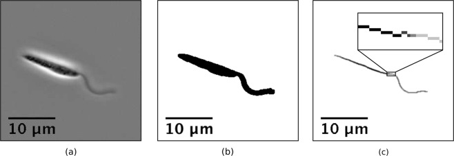
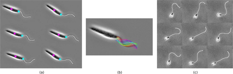
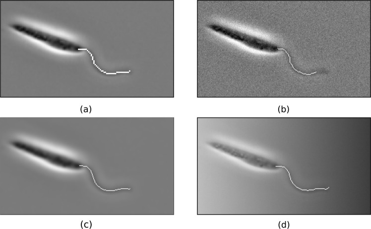
Ubiquitous in eukaryotic organisms, the flagellum is a well-studied organelle that is well-known to be responsible for motility in a variety of organisms. Commonly necessitated in their study is the capability to image and subsequently track the movement of one or more flagella using videomicroscopy, requiring digital isolation and location of the flagellum within a sequence of frames. Such a process in general currently requires some researcher input, providing some manual estimate or reliance on an experiment-specific heuristic to correctly identify and track the motion of a flagellum. Here we present a fully-automated method of flagellum identification from videomicroscopy based on the fact that the flagella are of approximately constant width when viewed by microscopy. We demonstrate the effectiveness of the algorithm by application to captured videomicroscopy of Leishmania mexicana, a parasitic monoflagellate of the family Trypanosomatidae. ImageJ Macros for flagellar identification are provided, and high accuracy and remarkable throughput are achieved via this unsupervised method, obtaining results comparable in quality to previous studies of closely-related species but achieved without the need for precursory measurements or the development of a specialised heuristic, enabling in general the automated generation of digitised kinematic descriptions of flagellar beating from videomicroscopy.


Conflict of interest statement

The authors declare no competing interests.

Figures

Figure 1
Figure 1
A sample frame taken from phase contrast videomicroscopy of a L. mexicana promastigote, from the dataset of Walker et al.. (a) Original frame. Despite the presence of an accessory structure, the paraflagellar rod, at the recorded resolution the flagellum appears to be of approximately-constant width. (b) Result of processing (a) into a binary image, following background subtraction and noise reduction. Existing methods of flagellum extraction are unable to automatically identify the flagellum in this image, typically requiring user input at this stage. (c) Result of the medial axis transform applied to (b), encoding the width of the cell along the medial line. Shown inset is an enlarged section of the transform.
Figure 2
Figure 2
Examples of the medial axis transform applied to low-resolution simple shapes. The original image is shown in black, with the results of the transform superimposed in greyscale, where brighter pixels correspond to higher values of the transform and thus wider sections of the original shape. (a) A simple disk is mapped to a single point by the transform, with the radius of the disk encoded in the transform. (b) A triangular region is reduced to the skeleton shown, with the encoded width decreasing away from the skeleton and triangle’s shared centre. (c) An annulus is transformed to a circle along its medial line, with a constant width-profile subject to artefacts of the rasterisation.
Figure 3
Figure 3
Analysis of a sample medial axis transform, corresponding to the cell in Fig. 1. (a) Values taken by the medial axis transform, shown in pixels, against the arclength, measured from left to right in Fig. 1. The flagellum may be clearly identified from this width-profile as the segment with approximately constant width, in contrast to the varied size of the rest of the cell. (b) A histogram of the discrete values of the transform shown in (a). A clear modal width can be seen around the flagellum width, suggesting that simple identification of the modal pixels may be sufficient to identify the flagellum in cases where the flagellar lengthscale is dominant.
Figure 4
Figure 4
Composite data and results from an implementation of the proposed scheme. A montage of original frames with the identified flagella superimposed, showing very good agreement with identification by eye, for sample (a,b) L. mexicana and (c) spermatozoa. In (a) the computed cell centroids and locations of flagellar attachment are marked as magenta squares and cyan circles respectively. In (b) captured flagella from multiple frames are superimposed on the first frame of the sample dataset, with the simple sinusoidal nature of the flagellar beating being clearly visible. Original frames from the datasets of Walker et al. (unpublished) and Ishimoto et al. respectively. Reprinted original frames of (b) with permission from [K. Ishimoto, H. Gadêlha, E.A. Gaffney, D.J. Smith, J. Kirkman-Brown. Physical Review Letters 118, 124501, 2017] Copyright (2017) by the American Physical Society.
Figure 5
Figure 5
Composite degraded data and results from an implementation of the proposed scheme, where identified flagella are shown superimposed. Data has been degraded by application of: (a) downsampling (2x); (b) Gaussian noise; (c) Gaussian blur; (d) pixel-wise multiplication by a black-to-white horizontal gradient. Acceptable flagellar segmentation by Macro 1 of the Supplementary Material is seen for each degraded dataset shown here, with some loss of accuracy at the tip or base of the flagellum. In particular, Gaussian noise is seen in this case to prevent the distal tip of the flagellum being segmented from the image background, a result of the basic preprocessing implemented here and not characteristic in general of the proposed flagellum segmentation procedure.

Similar articles

Cited by

References

    1. Scharer L, Littlewood DTJ, Waeschenbach A, Yoshida W, Vizoso DB. Mating behavior and the evolution of sperm design. Proc. Natl. Acad. Sci. USA. 2011;108:1490–1495. doi: 10.1073/pnas.1013892108. - DOI - PMC - PubMed
    1. Manton I, Clarke B. An electron microscope study of the spermatozoid of Sphagnum. J. Exp. Bot. 1952;3:265–275. doi: 10.1093/jxb/3.3.265. - DOI
    1. Fawcett DW. The mammalian spermatozoon. Dev. Biol. 1975;44:394–436. doi: 10.1016/0012-1606(75)90411-X. - DOI - PubMed
    1. de Souza W, Souto-Padrón T. The paraxial structure of the flagellum of Trypanosomatidae. J. Parasitol. 1980;66:229. doi: 10.2307/3280809. - DOI - PubMed
    1. Holwill ME, McGregor JL. Micromanipulation of the flagellum of Crithidia oncopelti. I. Mechanical effects. J. Exp. Biol. 1974;60:437–44. - PubMed

Publication types