. 2017;50(15):2412-2425.
doi: 10.1080/00032719.2017.1287713. Epub 2017 Sep 12.

VARIABLE SELECTION AND BIOMARKER CORRELATION IN THE ANALYSIS OF MYCOPLASMA PNEUMONIAE STRAINS BY SURFACE-ENHANCED RAMAN SPECTROSCOPY

Affiliations

VARIABLE SELECTION AND BIOMARKER CORRELATION IN THE ANALYSIS OF MYCOPLASMA PNEUMONIAE STRAINS BY SURFACE-ENHANCED RAMAN SPECTROSCOPY

Duncan C Krause et al. Anal Lett. 2017.

Abstract

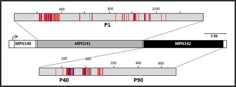
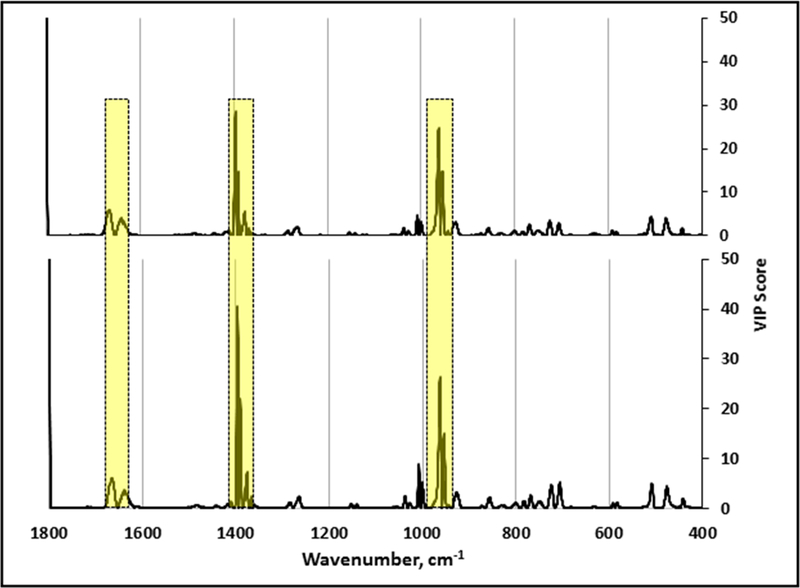
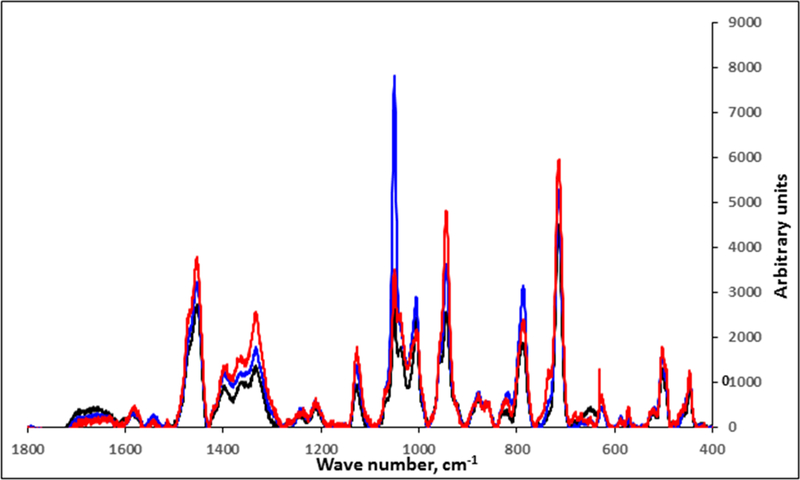
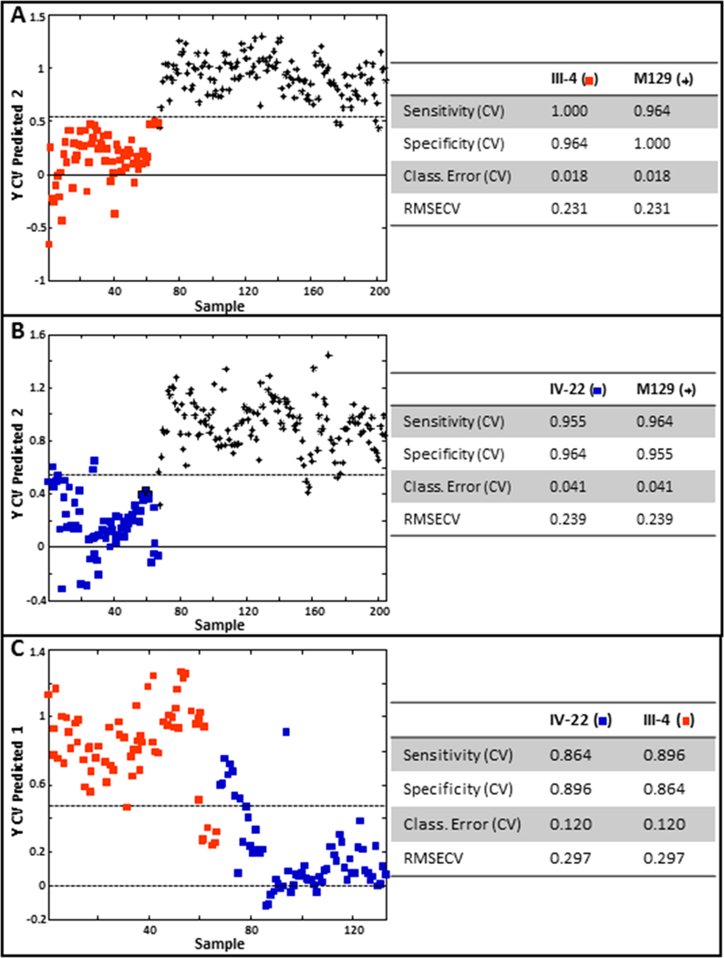
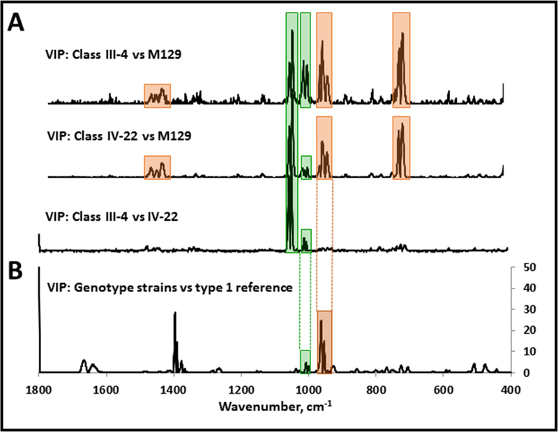
Mycoplasma pneumoniae is a human respiratory tract pathogen causing chronic bronchitis and atypical or "walking" pneumonia. The major surface protein P1 must form complexes with proteins P30 and P40/P90 in order to function in receptor binding and gliding motility, and variability in P1 and P40/P90 distinguishes the two major M. pneumoniae genotypes. Strains belonging to each genotype can be differentiated with high sensitivity and specificity by utilizing surface-enhanced Raman spectroscopy on silver nanorod arrays. Here we used the variable selection method of Variable Importance in Projection (VIP) to identify Raman bands important in M. pneumoniae strain classification. Furthermore, VIP analysis of mutants lacking P40/P90, or P1and P40/P90, correlated certain Raman bands important in distinguishing genotypes, with specific mycoplasma surface protein composition and presentation. Variable selection, and its correlation with specific mycoplasma surface components, is an important next step in developing this platform for M. pneumoniae detection and genotyping.

Keywords: Mycoplasma pneumoniae; Variable Importance in Projection; genotyping; nanorod array; surface-enhanced Raman spectroscopy.


Figures

Figure 1.
Figure 1.
Schematic representation of amino acid sequence variability in M. pneumoniae proteins P1 and P40/90 between the genotype 1 reference strain M129 and the genotype 2 reference strain FH. P1 and P40/P90 are the products of MPN141 and MPN142, respectively. Red green, and blue lines indicate amino acid substitutions, insertions, and deletions, respectively, in FH relative to M129.
Figure 2.
Figure 2.
VIP analysis of PLS-DA models that discriminate diverse clinical isolates relative to the genotype 1 reference strain M129 (upper; n= 195 spectra) or the genotype 2 reference strain FH (lower; n=175 spectra). Regions highlighted by yellow boxes indicate major discriminating Raman band wavenumbers. The band at wavenumber 1800 cm-1 was variable in mycoplasma samples and growth medium controls and is likely a function of sample preparation. As there are no known biomolecules assigned to this wavenumber, this band was not considered further here.
Figure 3.
Figure 3.
Baseline-corrected and averaged NA-SERS spectra for wild-type M129 (black; n=75) and mutants III-4 (red; n=68) and IV-22 (blue; n=67).
Figure 4.
Figure 4.
PLS-DA of mutant III-4 (A) or mutant IV-22 (B) relative to the wild-type parent strain M129, and of mutant III-4 relative to mutant IV-22. Stars, M129; red squares, mutant III-4; blue squares, mutant IV-22. Cross-validated statistics were obtained using Venetian blinds with 10 data splits to represent the prediction performance for each model. CV, cross-validated; RMSECV, root mean square error, cross-validated.
Figure 5.
Figure 5.
VIP analysis from pairwise PLS-DA modeling of (A) mutant III-4 with M129, mutant IV-22 with M129, and mutant III-4 with mutant IV-22, or (B) diverse clinical isolates relative to the genotype 1 reference strain M129. Orange, major band wavenumbers in common for VIP analysis of wild-type M129 and either mutant (A) and also from the genotype VIP analysis (B); green, major band wavenumbers in common for VIP analysis of wild-type M129 and either mutant, as well as VIP analysis of the two mutants (A) and also for the genotype VIP analysis (B).

References

    1. Atkinson TP, and Waites KB 2014. Mycoplasma pneumoniae infections in childhood. Pediatr. Inf. Dis. J. 33: 92–94. - PubMed
    1. Balish MF 2014. Mycoplasma pneumoniae, an underutilized model for bacterial cell biology. J. Bacteriol. 196: 3675–3682. - PMC - PubMed
    1. Ballabio D and Consonni V 2013. Classification tools in chemistry. Part 1: linear models. PLS-DA. Anal. Methods 5: 3790–3798.
    1. Barker M and Rayens W 2003. Partial least squares for discrimination. J. Chemometr. 17: 166–173.
    1. Baseman JB, Cole RM, Krause DC, and Leith DK 1982. Molecular basis for cytadsorption of Mycoplasma pneumoniae. J. Bacteriol 151: 1514–1522. - PMC - PubMed

LinkOut - more resources