

Anatomical study of the skull of amphisbaenian Diplometopon zarudnyi (Squamata, Amphisbaenia)
- PMID: 30899165
- PMCID: PMC6408683
- DOI: 10.1016/j.sjbs.2017.07.011
Anatomical study of the skull of amphisbaenian Diplometopon zarudnyi (Squamata, Amphisbaenia)
Abstract
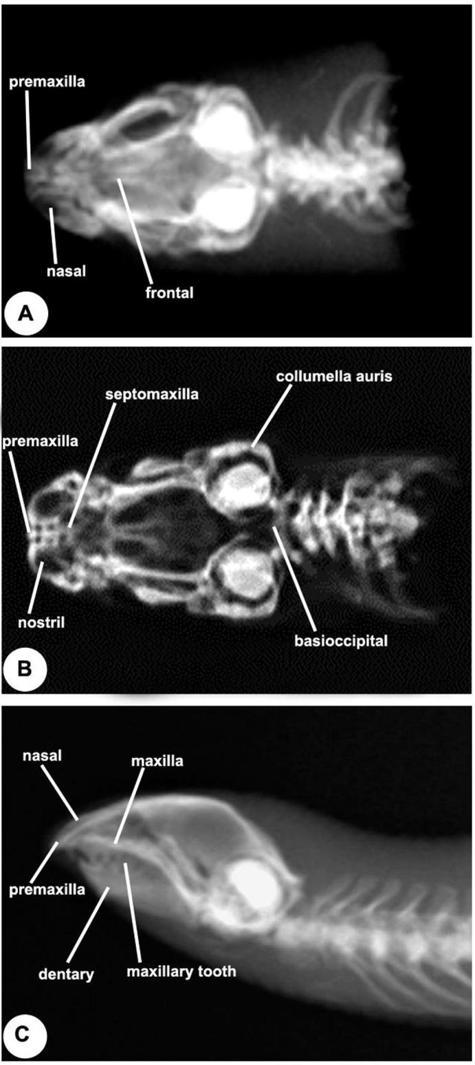
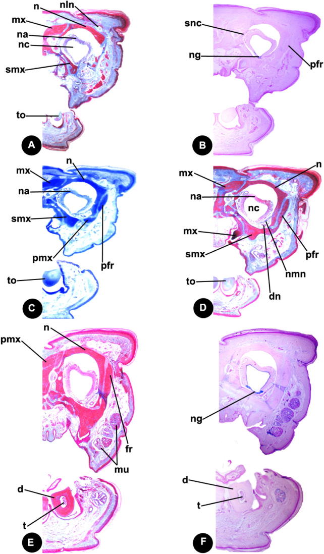
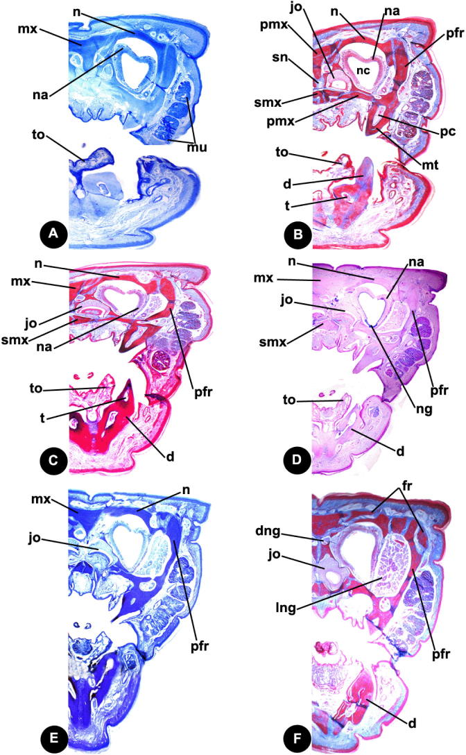
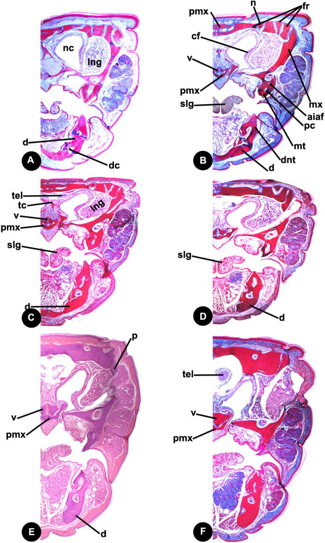
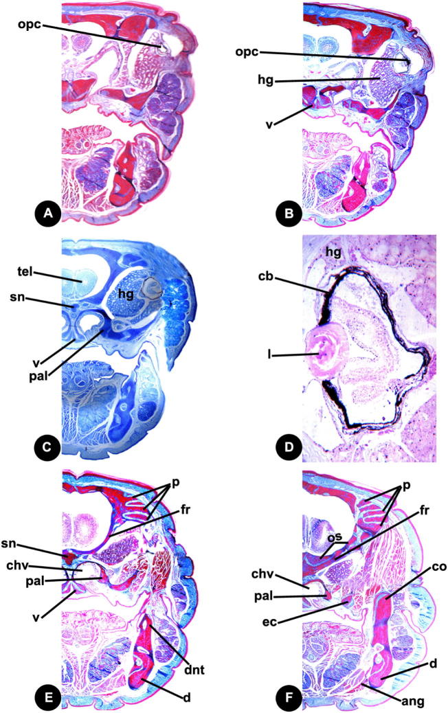
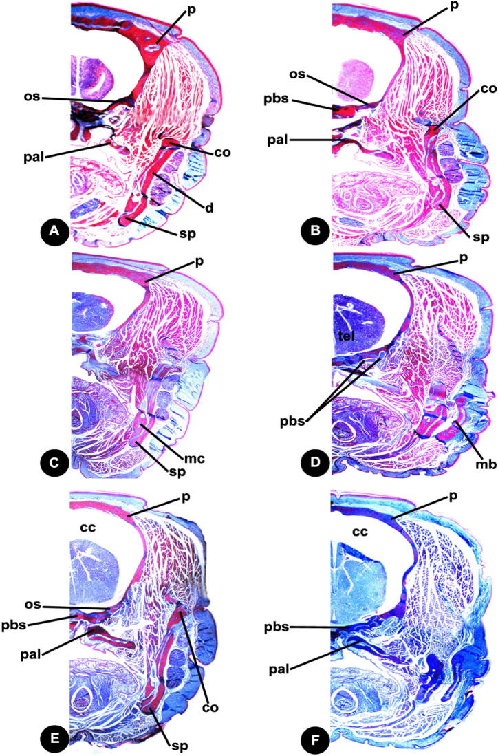
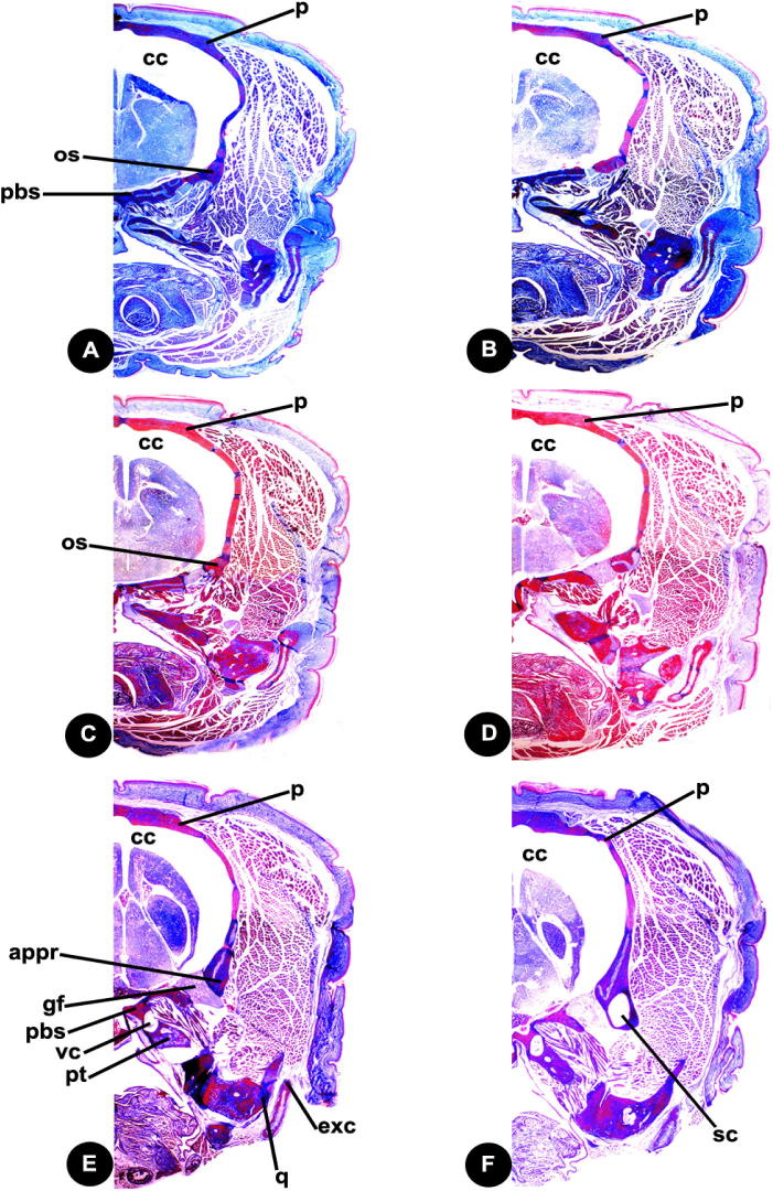
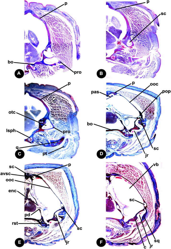
This study investigates the amphisbaenian species skull which includes cranium, lower jaw and hyoid apparatus. The medial dorsal bones comprise the premaxilla, nasal, frontal and parietal. The premaxilla carries a large medial tooth and two lateral ones. The nasals are paired bones and separated by longitudinal suture. Bones of circumorbital series are frontal, orbitosphenoid and maxilla. The occipital ring consists of basioccipital, supraoccipital and exooccipital. Supraoccipital and basioccipital are single bones while the exo-occipitals are paired. The bones of the palate comprise premaxilla, maxilla, septomaxilla, palatine, pterygoid, ectopterygoid, basisphenoid, parasphenoid, orbitosphenoid and laterosphenoid. Prevomer and pterygoid teeth are absent. Palatine represent by two separate bones. The temporal bones are clearly visible. The lower jaw consists of the dentary, articular, coronoid, supra-angular, angular and splenial. The hyoid apparatus is represented by a Y-shaped structure. The mandible is long and is suspended from the braincase via relatively short quadrate. There is an extensive contact between the long angular and the large triangular coronoid. Thus inter-mandibular joint is bridged completely by the angular and consequently, the lower jaws are relatively rigid and kinetic. The maxillae are suspended from the braincase largely by ligaments and muscles rather than through bony articulation. In conclusion, the skull shape affects feeding strategy in Diplometopon zarudnyi. The prey is ingested and transported via a rapid maxillary raking mechanism.
Keywords: Amphisbaenia; Diplometopon zarudnyi; Morphology; Skull; Squamata.
Figures
References
-
- Abdeen A.M. Further studies on the ophidian cranial osteology: the skull of the Egyptian black cobra, Walterinnesia aegyptia, (Family Elapidae). A. The dorsal bones, bones of the upper jaw, circumorbital series and occipital ring. J. Egypt Ger. Soc. Zool. 1991;5:367–389.
-
- Abdeen A.M., Zaher M.M. Futher studies on the ophidian osteocrania: the osteocranium of the snake Eryx jaculus (Family: Boidae) J. Egypt Ger. Soc. Zool. 1992;7(B):295–333.
-
- Abdeen A.M., Abo-Taira A.M., Zaher M.M. Further studies on the Ophidian cranial osteology: the skull of the Egyptian blind snake Leptotyphlops cairi (Family, Leptotyphlopidae) I. The cranium. A-The medial dorsal bones, bones of the upper jaw, circumorbital series and occipital ring. J. Egypt Ger. Soc. Zool. 1991;5:417–437.
-
- Abdeen A.M., Abo-Taira A.M., Zaher M.M. Further studies on the Ophidian cranial osteology: the skull of the Egyptian blind snake Leptotyphlops cairi (Family, Leptotyphlopidae) I. The cranium. B-The otic capsule, palate and temporal bones. J. Egypt Ger. Soc. Zool. 1991;5:439–455.
-
- Allam A.A., Abo El–Eneen R.E. Scales microstructure of some snakes inhabited the Egyptian Area. Zool. Sci. 2012;29(11):770–775. - PubMed
LinkOut - more resources
Full Text Sources
