

Alterations in GABAA-Receptor Trafficking and Synaptic Dysfunction in Brain Disorders
- PMID: 30899215
- PMCID: PMC6416223
- DOI: 10.3389/fncel.2019.00077
Alterations in GABAA-Receptor Trafficking and Synaptic Dysfunction in Brain Disorders
Abstract
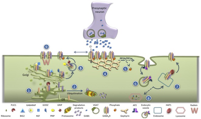
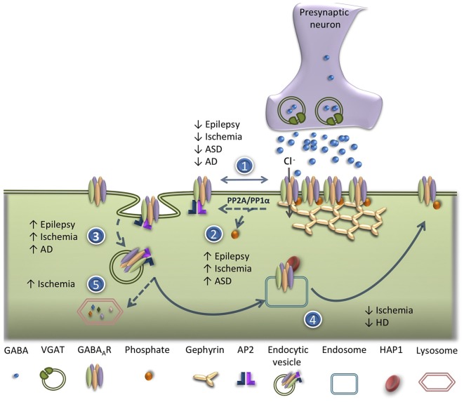
GABAA receptors (GABAAR) are the major players in fast inhibitory neurotransmission in the central nervous system (CNS). Regulation of GABAAR trafficking and the control of their surface expression play important roles in the modulation of the strength of synaptic inhibition. Different pieces of evidence show that alterations in the surface distribution of GABAAR and dysregulation of their turnover impair the activity of inhibitory synapses. A diminished efficacy of inhibitory neurotransmission affects the excitatory/inhibitory balance and is a common feature of various disorders of the CNS characterized by an increased excitability of neuronal networks. The synaptic pool of GABAAR is mainly controlled through regulation of internalization, recycling and lateral diffusion of the receptors. Under physiological condition these mechanisms are finely coordinated to define the strength of GABAergic synapses. In this review article, we focus on the alteration in GABAAR trafficking with an impact on the function of inhibitory synapses in various disorders of the CNS. In particular we discuss how similar molecular mechanisms affecting the synaptic distribution of GABAAR and consequently the excitatory/inhibitory balance may be associated with a wide diversity of pathologies of the CNS, from psychiatric disorders to acute alterations leading to neuronal death. A better understanding of the cellular and molecular mechanisms that contribute to the impairment of GABAergic neurotransmission in these disorders, in particular the alterations in GABAAR trafficking and surface distribution, may lead to the identification of new pharmacological targets and to the development of novel therapeutic strategies.
Keywords: Alzheimer’s disease; GABAA receptor trafficking; Huntington’s disease; Parkinson’s disease; brain ischemia; epilepsy.
Figures
Similar articles
-
Role of GABAA R trafficking in the plasticity of inhibitory synapses.J Neurochem. 2016 Dec;139(6):997-1018. doi: 10.1111/jnc.13742. Epub 2016 Sep 30. J Neurochem. 2016. PMID: 27424566 Review.
-
Interaction of different proteins with GABAA receptor and their modulatory effect on inhibitory neural transmission leads to epilepsy.CNS Neurol Disord Drug Targets. 2014;13(7):1148-59. doi: 10.2174/1871527313666140917115121. CNS Neurol Disord Drug Targets. 2014. PMID: 25230226 Review.
-
Clptm1 Limits Forward Trafficking of GABAA Receptors to Scale Inhibitory Synaptic Strength.Neuron. 2018 Feb 7;97(3):596-610.e8. doi: 10.1016/j.neuron.2017.12.038. Epub 2018 Jan 25. Neuron. 2018. PMID: 29395912 Free PMC article.
-
γ2 GABAAR Trafficking and the Consequences of Human Genetic Variation.Front Cell Neurosci. 2018 Aug 23;12:265. doi: 10.3389/fncel.2018.00265. eCollection 2018. Front Cell Neurosci. 2018. PMID: 30190672 Free PMC article. Review.
-
Downregulation of GABAA Receptor Recycling Mediated by HAP1 Contributes to Neuronal Death in In Vitro Brain Ischemia.Mol Neurobiol. 2017 Jan;54(1):45-57. doi: 10.1007/s12035-015-9661-9. Epub 2016 Jan 5. Mol Neurobiol. 2017. PMID: 26732589
Cited by
-
Neuronal Transmembrane Chloride Transport Has a Time-Dependent Influence on Survival of Hippocampal Cultures to Oxygen-Glucose Deprivation.Brain Sci. 2019 Dec 6;9(12):360. doi: 10.3390/brainsci9120360. Brain Sci. 2019. PMID: 31817665 Free PMC article.
-
The Yin and Yang of GABAergic and Glutamatergic Synaptic Plasticity: Opposites in Balance by Crosstalking Mechanisms.Front Synaptic Neurosci. 2022 May 19;14:911020. doi: 10.3389/fnsyn.2022.911020. eCollection 2022. Front Synaptic Neurosci. 2022. PMID: 35663370 Free PMC article. Review.
-
GABAA Receptor β3 Subunit Mutation N328D Heterozygous Knock-in Mice Have Lennox-Gastaut Syndrome.Int J Mol Sci. 2023 May 8;24(9):8458. doi: 10.3390/ijms24098458. Int J Mol Sci. 2023. PMID: 37176165 Free PMC article.
-
The role of GABA in islet function.Front Endocrinol (Lausanne). 2022 Sep 29;13:972115. doi: 10.3389/fendo.2022.972115. eCollection 2022. Front Endocrinol (Lausanne). 2022. PMID: 36246925 Free PMC article. Review.
-
Forskolin reverses the O-GlcNAcylation dependent decrease in GABAAR current amplitude at hippocampal synapses possibly at a neurosteroid site on GABAARs.Res Sq [Preprint]. 2024 Apr 10:rs.3.rs-4140038. doi: 10.21203/rs.3.rs-4140038/v1. Res Sq. 2024. Update in: Sci Rep. 2024 Jul 29;14(1):17461. doi: 10.1038/s41598-024-66025-w. PMID: 38659738 Free PMC article. Updated. Preprint.
References
-
- Baio J., Wiggins L., Christensen D. L., Maenner M. J., Daniels J., Warren Z., et al. . (2018). Prevalence of autism spectrum disorder among children aged 8 years—autism and developmental disabilities monitoring network, 11 sites, united states, 2014. MMWR Surveill. Summ. 67, 1–23. 10.15585/mmwr.ss6706a1 - DOI - PMC - PubMed
LinkOut - more resources
Full Text Sources