. 2019 Mar 7:10:339.
doi: 10.3389/fimmu.2019.00339. eCollection 2019.

CXCR6-Deficiency Improves the Control of Pulmonary Mycobacterium tuberculosis and Influenza Infection Independent of T-Lymphocyte Recruitment to the Lungs

Affiliations

CXCR6-Deficiency Improves the Control of Pulmonary Mycobacterium tuberculosis and Influenza Infection Independent of T-Lymphocyte Recruitment to the Lungs

Anneliese S Ashhurst et al. Front Immunol. .

Abstract

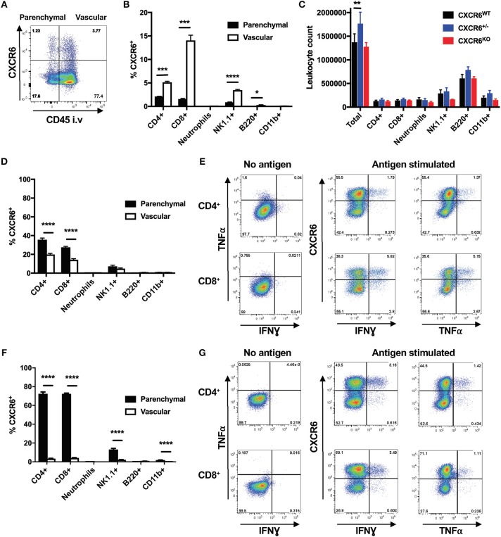
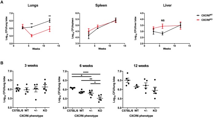
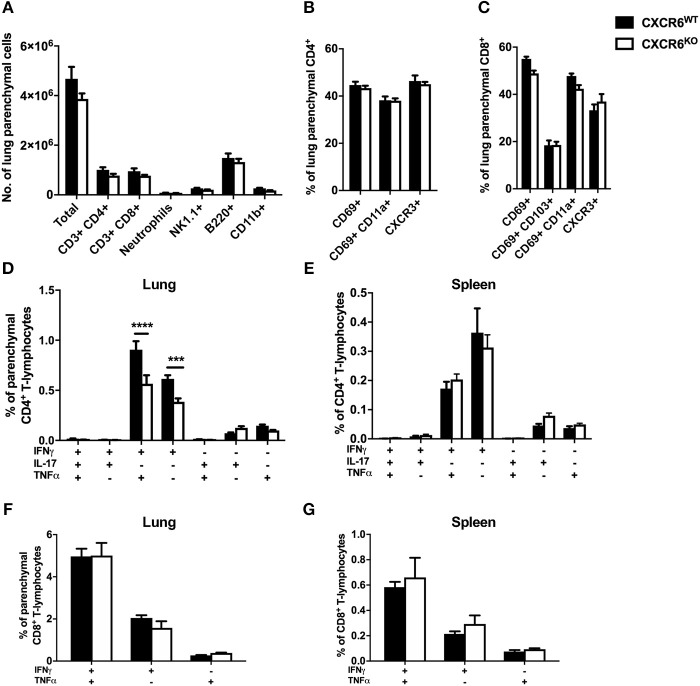
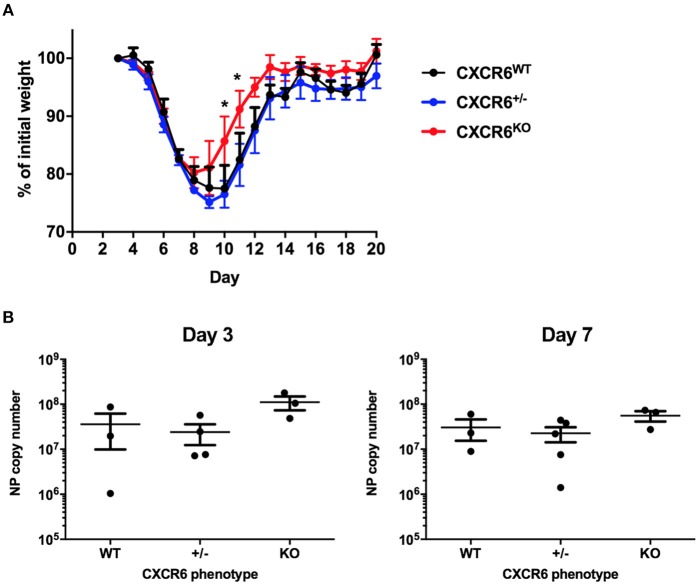
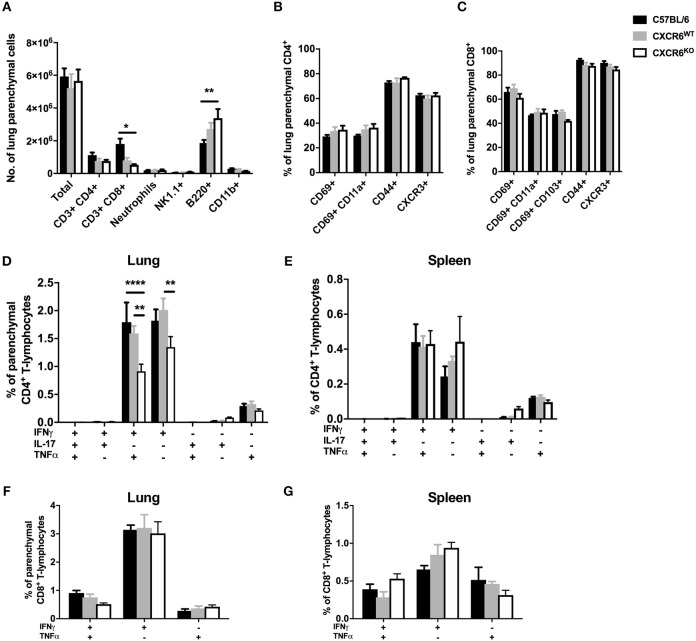
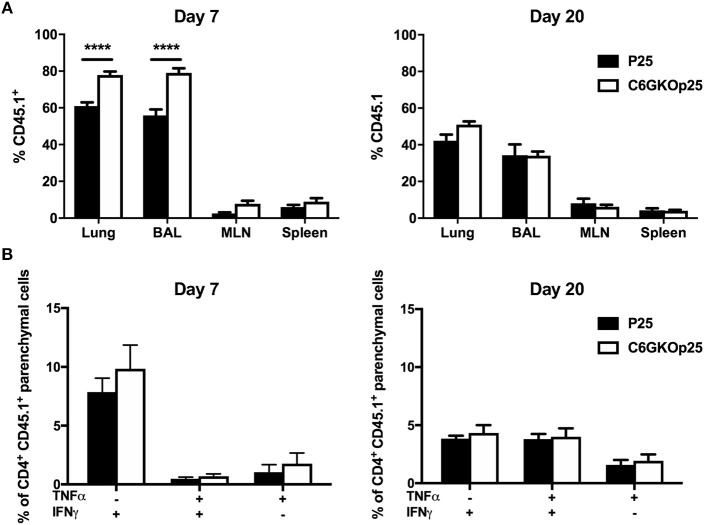
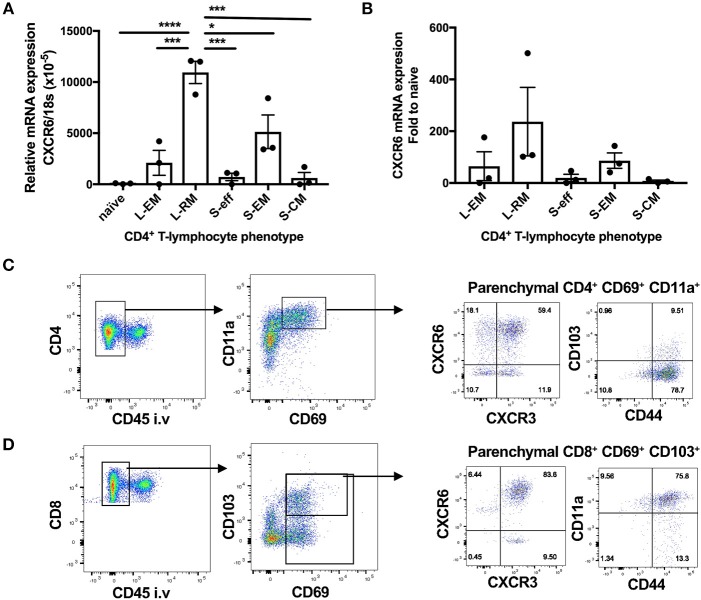
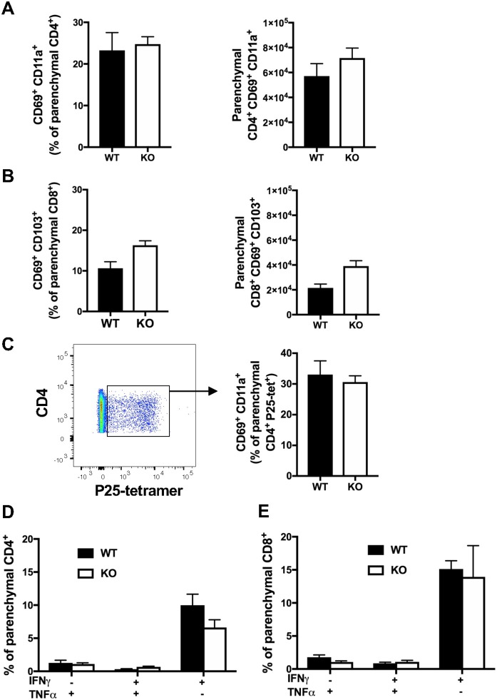
T-lymphocytes are critical for protection against respiratory infections, such as Mycobacterium tuberculosis and influenza virus, with chemokine receptors playing an important role in directing these cells to the lungs. CXCR6 is expressed by activated T-lymphocytes and its ligand, CXCL16, is constitutively expressed by the bronchial epithelia, suggesting a role in T-lymphocyte recruitment and retention. However, it is unknown whether CXCR6 is required in responses to pulmonary infection, particularly on CD4+ T-lymphocytes. Analysis of CXCR6-reporter mice revealed that in naïve mice, lung leukocyte expression of CXCR6 was largely restricted to a small population of T-lymphocytes, but this population was highly upregulated after either infection. Nevertheless, pulmonary infection of CXCR6-deficient mice with M. tuberculosis or recombinant influenza A virus expressing P25 peptide (rIAV-P25), an I-Ab-restricted epitope from the immunodominant mycobacterial antigen, Ag85B, demonstrated that the receptor was redundant for recruitment of T-lymphocytes to the lungs. Interestingly, CXCR6-deficiency resulted in reduced bacterial burden in the lungs 6 weeks after M. tuberculosis infection, and reduced weight loss after rIAV-P25 infection compared to wild type controls. This was paradoxically associated with a decrease in Th1-cytokine responses in the lung parenchyma. Adoptive transfer of P25-specific CXCR6-deficient T-lymphocytes into WT mice revealed that this functional change in Th1-cytokine production was not due to a T-lymphocyte intrinsic mechanism. Moreover, there was no reduction in the number or function of CD4+ and CD8+ tissue resident memory cells in the lungs of CXCR6-deficient mice. Although CXCR6 was not required for T-lymphocyte recruitment or retention in the lungs, CXCR6 influenced the kinetics of the inflammatory response so that deficiency led to increased host control of M. tuberculosis and influenza virus.

Keywords: CXCR6; influenza; lung; tissue-resident memory; tuberculosis.


Figures

Figure 1
Figure 1
Expression pattern of CXCR6 in the lungs of mice. CXCR6-reporter mice were injected i.v with anti-CD45 antibody to label intra-vascular lung leukocytes immediately prior to collection of lung cells and analysis by flow cytometry. (A) CXCR6 or CXCR6+ leukocyte populations in the lung parenchyma (CD45) or vasculature (CD45+) of naïve mice. (B) Percentage of leukocyte populations in naïve lung parenchyma or vasculature expressing CXCR6 (n = 3). (C) Enumeration of different leukocyte populations in the lungs of naïve CXCR6WT, CXCR6+/−, and CXCR6KO mice (n = 3). (D,E) CXCR6-reporter mice (n = 5) were infected with M. tuberculosis (~100 CFU) for 6 weeks. (D) Percentage of different leukocyte populations in the lung parenchyma or vasculature expressing CXCR6. (E) Lymphocytes from the lungs were recalled with peptide antigen, or as a control were cultured in the same manner but without antigen, and the expression of CXCR6 by parenchymal cytokine-secreting P25-specific CD4+ and TB10.43−11-specific CD8+ T-lymphocytes was determined. (F,G) CXCR6-reporter mice (n = 4) were infected i.n with PR8-P25. (F) Percentage of leukocyte populations in the lung parenchyma or vasculature expressing CXCR6 at day 10. (G) Lymphocytes from the lungs at day 20 were recalled with peptide antigen, or as a control were cultured in the same manner but without antigen, and the expression of CXCR6 by parenchymal cytokine-secreting P25-specific CD4+ and NP366−375-specific CD8+ T-lymphocytes was determined. Data are the means ± SEM and are representative of repeat experiments. The statistical significance of differences were analyzed by (B,D,F) multiple t-tests with correction for multiple comparisons using the Holm-Sidak method or (C) by ANOVA with Dunnett's multiple comparisons test (*p < 0.05, **p < 0.01, ***p < 0.001, ****p < 0.0001).
Figure 2
Figure 2
CXCR6-deficient mice have improved control of M. tuberculosis infection in the lungs. (A) CXCR6WT (C57BL/6, black line) or CXCR6KO (red line) mice (n = 5) were infected with M. tuberculosis (~100 CFU) by aerosol, and the kinetics of bacterial growth in the lungs, spleen, and liver were determined at 3, 6, and 12 weeks. (B) C57BL/6 and littermate matched CXCR6WT, CXCR6+/−, and CXCR6KO mice (n = 3–5) were similarly infected and bacterial counts enumerated in the lungs at 3, 6, and 12 weeks. Data are the means ± SEM and are representative of four experiments. The statistical significance of differences between groups and WT controls were analyzed by ANOVA with Bonferroni post-hoc comparison (*p < 0.05, **p < 0.01, ****p < 0.0001, NS, not significant).
Figure 3
Figure 3
CXCR6-deficient mice have reduced Th1 T-lymphocyte responses in the lung parenchyma without changes to leukocyte recruitment at 6 weeks of M. tuberculosis infection. CXCR6WT (C57BL6; closed bars) or CXCR6KO (open bars) mice (n = 5) were infected with M. tuberculosis by aerosol and at 6 weeks were injected i.v with anti-CD45 antibody to label intra-vascular leukocytes. (A) Leukocyte recruitment to the lung parenchyma (CD45) was enumerated by flow cytometry. Expression of surface activation markers and CXCR3 was assessed on (B) CD4+ and (C) CD8+ T-lymphocytes. Leukocytes were stimulated with peptide followed by ICS and flow cytometry. P25-specific CD4+ T-lymphocyte cytokine expression in the (D) lung parenchyma (CD45) and (E) spleen. TB10.43−11-specific CD8+ T-lymphocyte cytokine expression in the (F) lung parenchyma (CD45) and (G) spleen. Data are the means ± SEM and are representative of three repeat experiments. The statistical significance of differences between groups were analyzed by ANOVA with Bonferroni post-hoc comparison (***p < 0.001, ****p < 0.0001).
Figure 4
Figure 4
CXCR6-deficient mice experience reduced weight loss after acute influenza infection without change in viral load. (A) Comparison of weight loss after i.n PR8-P25 infection between CXCR6WT, CXCR6+/−, CXCR6KO littermates, as a percentage of weight for individual mice from day 3 (n = 4–5). (B) NP viral copy number was enumerated in the lungs at days 3 and 7 after infection by RT-PCR in littermates (n = 3–5). Data are the means ± SEM and are representative of repeat experiments. The statistical significance of differences between groups were analyzed by ANOVA with Bonferroni post-hoc comparison to WT controls (*p < 0.05).
Figure 5
Figure 5
CXCR6-deficient mice have reduced Th1 T-lymphocyte responses in the lung parenchyma at day 20 after influenza infection without changes to leukocyte recruitment. C57BL/6 (closed bars), CXCR6WT (gray bars), or CXCR6KO (open bars) mice (n = 4) were infected with PR8-P25 and at 20 days were injected i.v with anti-CD45 antibody to label intra-vascular leukocytes. (A) Leukocyte recruitment to the lung parenchyma (CD45) was enumerated by flow cytometry. Expression of surface activation markers and CXCR3 was assessed on (B) CD4+ and (C) CD8+ T-lymphocytes. Leukocytes were stimulated with peptide followed by ICS and flow cytometry. P25-specific CD4+ T-lymphocyte cytokine expression in the (D) lung parenchyma (CD45) and (E) spleen. NP366−375-specific CD8+ T-lymphocyte cytokine expression in the (F) lung parenchyma (CD45) and (G) spleen. Data are the means ± SEM and are representative of repeat experiments. The statistical significance of differences were analyzed by ANOVA with Bonferroni post-hoc comparison (*p < 0.05, **p < 0.01, ****p < 0.0001).
Figure 6
Figure 6
CXCR6-deficient CD4+ T-lymphocytes are recruited to the lung parenchyma and airways more rapidly than WT cells, but have comparable Th1-type cytokine responses. Female C57BL/6 mice (n = 4) received 5 × 10⁴ wild type (P25) or CXCR6KO P25-specific (C6GKO-P25) CD45.1+ CD4+ T-lymphocytes by adoptive transfer and 1 day later were infected i.n with PR8-P25. At 7 or 20 days, infected mice were injected i.v with anti-CD45 antibody to label intra-vascular leukocytes. (A) Proportion of total CD4+ T-lymphocytes in the lung parenchyma, BAL, MLN, and spleen that were CD45.1+. (B) Lung leukocytes were stimulated with P25 peptide and cytokine expression assessed in CD4+ CD45.1+ lung parenchymal cells by intra-cellular staining. Data are the means ± SEM and are representative of repeat experiments. The statistical significance of differences between groups were analyzed by ANOVA with Bonferroni post-hoc comparison (****p < 0.0001).
Figure 7
Figure 7
CXCR6 is highly expressed by lung resident memory T-lymphocytes after influenza infection. (A,B) 6 weeks following transfer of P25-specific CD45.1+ CD4+ T-lymphocytes (naïve) and infection with PR8-P25, transferred cells were purified from the lungs, and spleens by sorting according to memory phenotype: lung effector memory (L-EM, CD69CD62L), lung resident memory (L-RM, CD69+), spleen effector memory (S-EM, CD69CD62L), or spleen central memory (S-CM, CD69CD62L+). Effector P25 cells (S-eff) were sorted at 11 days p.i. The transcriptional expression of CXCR6 was determined by RT-PCR. Data are the means ± SEM of three replicate experiments (total n = 20) and are shown as (A) CXCR6 expression relative to 18S mRNA and (B) fold change in CXCR6 expression compared to naïve cells. The statistical significance of differences were analyzed by ANOVA with Bonferroni post-hoc comparison (*p < 0.05, ***p < 0.001, ****p < 0.0001). (C,D) CXCR6-reporter mice (n = 5) were infected i.n with PR8-P25 and at 6 weeks were injected i.v with anti-CD45 antibody to label intra-vascular leukocytes. Memory T-lymphocytes were identified by flow cytometry. Identification of (C) CD4+ TRM [parenchymal (CD45) CD69+ CD11a+] and (D) CD8+ TRM [parenchymal (CD45) CD69+ CD103+]. Expression of CXCR3, CXCR6, CD44, and CD103 are shown. Data are representative of repeat experiments.
Figure 8
Figure 8
CXCR6-deficiency does not impair the retention or function of tissue-resident T-lymphocytes in the lungs at 6 weeks. CXCR6WT or CXCR6KO mice (n = 5) were infected i.n with PR8-P25 and at 6 weeks were injected i.v with anti-CD45 antibody to label intra-vascular leukocytes. Endogenous memory T-lymphocytes were identified by flow cytometry. The proportion and number of total (A) CD4+ TRM [parenchymal (CD45) CD69+ CD11a+] and (B) CD8+ TRM [parenchymal (CD45) CD69+ CD103+]. (C) Proportion of P25-specific CD4+ TRM [parenchymal (CD45-) CD4+ P25-tetramer+ CD69+ CD11a+] were identified by tetramer staining. Lung leukocytes were incubated with (D) P25 peptide to stimulate memory CD4+ T-lymphocytes or (E) NP366−375 peptide to stimulate memory CD8+ T-lymphocytes, followed by ICS and flow cytometry to assess cytokine production by parenchymal cells (CD45). Data are the means ± SEM and are representative of repeat experiments. The statistical significance of differences were analyzed by ANOVA with Bonferroni post-hoc comparison.

Similar articles

Cited by

References

    1. Iijima N, Iwasaki A. A local macrophage chemokine network sustains protective tissue-resident memory CD4 T cells. Science. (2014) 346:93–8. 10.1126/science.1257530 - DOI - PMC - PubMed
    1. Tse S-W, Radtke AJ, Espinosa DA, Cockburn IA, Zavala F. The chemokine receptor CXCR6 is required for the maintenance of liver memory CD8+ T cells specific for infectious pathogens. J Infect Dis. (2014) 210:1508–16. 10.1093/infdis/jiu281 - DOI - PMC - PubMed
    1. Alkhatib G, Liao F, Berger EA, Farber JM, Peden KWC. A new SIV co-receptor, STRL33. Nature. (1997) 388:238. 10.1038/40789 - DOI - PubMed
    1. Kim CH, Kunkel EJ, Boisvert J, Johnston B, Campbell JJ, Genovese MC, et al. . Bonzo/CXCR6 expression defines type 1–polarized T-cell subsets with extralymphoid tissue homing potential. J Clin Invest. (2001) 107:595–601. 10.1172/JCI11902 - DOI - PMC - PubMed
    1. Deng L, Chen N, Li Y, Zheng H, Lei Q. CXCR6/CXCL16 functions as a regulator in metastasis and progression of cancer. Biochim Biophys Acta. (2010) 1806:42–9. 10.1016/j.bbcan.2010.01.004 - DOI - PubMed

Publication types

MeSH terms