. 2019 Mar 7:10:377.
doi: 10.3389/fimmu.2019.00377. eCollection 2019.

Inhibition of JAK1/2 Tyrosine Kinases Reduces Neurogenic Heterotopic Ossification After Spinal Cord Injury

Affiliations

Inhibition of JAK1/2 Tyrosine Kinases Reduces Neurogenic Heterotopic Ossification After Spinal Cord Injury

Kylie A Alexander et al. Front Immunol. .

Abstract

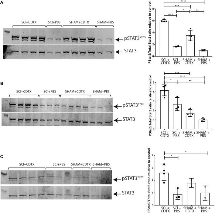
Neurogenic heterotopic ossifications (NHO) are very incapacitating complications of traumatic brain and spinal cord injuries (SCI) which manifest as abnormal formation of bone tissue in periarticular muscles. NHO are debilitating as they cause pain, partial or total joint ankylosis and vascular and nerve compression. NHO pathogenesis is unknown and the only effective treatment remains surgical resection, however once resected, NHO can re-occur. To further understand NHO pathogenesis, we developed the first animal model of NHO following SCI in genetically unmodified mice, which mimics most clinical features of NHO in patients. We have previously shown that the combination of (1) a central nervous system lesion (SCI) and (2) muscular damage (via an intramuscular injection of cardiotoxin) is required for NHO development. Furthermore, macrophages within the injured muscle play a critical role in driving NHO pathogenesis. More recently we demonstrated that macrophage-derived oncostatin M (OSM) is a key mediator of both human and mouse NHO. We now report that inflammatory monocytes infiltrate the injured muscles of SCI mice developing NHO at significantly higher levels compared to mice without SCI. Muscle infiltrating monocytes and neutrophils expressed OSM whereas mouse muscle satellite and interstitial cell expressed the OSM receptor (OSMR). In vitro recombinant mouse OSM induced tyrosine phosphorylation of the transcription factor STAT3, a downstream target of OSMR:gp130 signaling in muscle progenitor cells. As STAT3 is tyrosine phosphorylated by JAK1/2 tyrosine kinases downstream of OSMR:gp130, we demonstrated that the JAK1/2 tyrosine kinase inhibitor ruxolitinib blocked OSM driven STAT3 tyrosine phosphorylation in mouse muscle progenitor cells. We further demonstrated in vivo that STAT3 tyrosine phosphorylation was not only significantly higher but persisted for a longer duration in injured muscles of SCI mice developing NHO compared to mice with muscle injury without SCI. Finally, administration of ruxolitinib for 7 days post-surgery significantly reduced STAT3 phosphorylation in injured muscles in vivo as well as NHO volume at all analyzed time-points up to 3 weeks post-surgery. Our results identify the JAK/STAT3 signaling pathway as a potential therapeutic target to reduce NHO development following SCI.

Keywords: JAK- STAT signaling pathway; heterotopic ossification; oncostatin M receptor; ruxolitinib; spinal cord injury complications.


Figures

Figure 1
Figure 1
Increased inflammatory monocyte infiltration in injured muscles of mice developing NHO. (A) C57BL/6 mice underwent either SCI or Sham surgery followed by an intra muscular injection of CDTX or PBS. Muscle leukocytes were extracted from hamstrings at day 4 post-surgery and isolated into four subsets based on forward scatter, side scatter as well as zombie aqua negativity (viable cells), F4/80, Ly6G, CD11b, and Ly6C expression. (B) Frequencies of each leukocyte population relative to total live muscle cells using the gating strategy outlined: (i) “Total F4/80+ cells” (CD11b+F4/80+, blue gates in A), (ii) “Ly6Chi inflammatory monocytes” (F4/80+CD11b+Ly6G CD48+Ly6Chi, blue gates in A), (iii) “Ly6Cmid/lo monocytes/macrophages” (F4/80+ CD11b+ Ly6GCD48+ Ly6Cmid/lo, blue gates in A), and (iv) “granulocytes” (F4/80CD11b+Ly6G+, red gates in A). The frequency of Ly6Chi inflammatory monocytes (relative to total live muscle cells) in mice developing NHO (SCI+CDTX), compared to all other treatment groups was significantly higher (p = 0.0002 ANOVA n = 3–5/group). Each dot represents an individual mouse. Bars represent as mean ± SD. (C) Muscle leukocytes were extracted from hamstrings at day 4 post surgery, and 4 separate leukocyte populations were identified by flow cytometry using the following gating strategy: (i) “Ly6Chi inflammatory monocytes” as CD45+ lineage (Ter119,CD3ε,B220)-negative F4/80+ CD11b+ Ly6G CD48+Ly6Chi, red gates, (ii) “Ly6Cmid monocytes/macrophages” CD45+ Lin F4/80+ CD11b+ Ly6GCD48+ Ly6Cmid, red gates, (iii) “Ly6Cneg monocytes” CD45+ Lin F4/80+ CD11b+ Ly6G CD48+ Ly6Cneg red gates, and (iv) “granulocytes” CD45+ Lin F4/80neg CD11b+ Ly6G+, blue gates. (D) Frequencies of each myeloid subset relative to total live muscle cells and to total live CD45+ muscle leukocytes. There was a significant increase in frequency of Ly6Chi monocytes relative to total live muscle cells (Di, p < 0.0001 Mann-Whitney test) and to CD45+ live muscle leukocytes (Di, p < 0.0001 Mann-Whitney test) after SCI+CDTX compared to Sham+CDTX. Each dot represents an individual mouse, n = 25–27/treatment group. Bars represent mean ± SD.
Figure 2
Figure 2
OSM and OSMR expression and signaling in muscle cells. (A) Osm mRNA expression by qRT-PCR. Osm is expressed by all sorted infiltrating myeloid populations but absent in whole naïve skeletal muscle. Each dot represents an individual mouse (n = 3–4/group). Bars represent mean ± SD. (B) Osmr mRNA expression is only present in sorted mouse muscle satellite and interstitial cells, not in any sorted myeloid population infiltrating the injured muscle (C) Phos-flow of pSTAT3 Y705 phosphorylation in cultured CD45Ter119CD31CD34+Sca1+ muscle interstitial cells (IC), CD45Ter119CD31CD34+Sca1 muscle satellite cells (SC), and Kusa4b10 cells. Cells were incubated for 10 min at 37°C with medium alone (unstimulated), or 25 ng/ml recombinant mouse OSM plus DMSO or of 25 ng/ml OSM plus 1 μM ruxolitinib. Cell were then fixed and permeabilized before staining with AlexaFluor647-conjugated mouse mAb anti-pSTAT3 (Y705) and analyzed by flow cytometry. Representative figures were one of two independent experiments.
Figure 3
Figure 3
Persistence of STAT3 tyrosine phosphorylation in injured muscles following spinal cord injury. Western-blots of whole muscle lysates collected from mice that underwent either SCI or SHAM surgery together with an intramuscular injection of either CDTX or PBS. Western blot of whole muscle lysates from individual mice taken on day 4 (A), 7 (B), or 14 (C) post-injury. Each membrane was probed with rabbit anti-pSTAT3 Y705 mAb, stripped and then re-probed with rabbit anti-total STAT3 mAb. Band fluorescence intensity was quantified and ratio of signal intensity of pSTAT3 vs. total STAT3 calculated for each individual mouse and normalized relative to the average pSTAT3/STAT3 ratio in control mice (SHAM+PBS) at each time-point. Each lane and each dot represents a separate mouse, n = 2–4 mice/treatment/time-point. Bars represent mean ± SD. P values were calculated by ANOVA, *p < 0.05, **p < 0.01, ***p < 0.001, ****p < 0.0001.
Figure 4
Figure 4
Inhibition of JAK1/2 kinases with ruxolitinib reduces NHO development after SCI in vivo. (A) Western-blots of whole muscle lysates collected at day 7 from SCI+CDTX mice that were treated with either vehicle control or ruxolitinib (60 mg/kg bi-daily) from day 0–7 post surgery. Western-blots of whole muscle lysates were probed with rabbit anti-pSTAT3 Y705 mAb, and rabbit anti-total STAT3 mAb, then band fluorescence was quantified and ratio of signal intensity of pSTAT3 versus total STAT3 calculated for each individual mouse. Each lane and each dot represents a separate mouse, n = 4–5/treatment group. Data represented as mean ± SD, *p = 0.03 by Mann-Whitney test. (B) Measurement of NHO volume by micro CT (μCT) in mice which received SCI+CDTX and treated with either vehicle control or ruxolitinib (60 mg/kg bi-daily) from day 0–7 post surgery. (i) NHO volumes were quantified in vivo by μCT at indicated time points post-surgery illustrating the reduction in NHO development. Each dot represents a separate mouse, n = 4–10 mice/treatment/time point. Data represented as mean ± SD, **p = 0.0076, *p = 0.031, and p = 0.015 respectively by Mann-Whitney test. (ii) Representative μCT images at 7 days post-surgery (C) Masson's trichrome staining 3 weeks post-surgery confirming the development of multiple NHO bone and collagen+ foci (crosshatches) within the muscle in vehicle treated mice, which are reduced after ruxolitinib treatment, and absent in control mice (SHAM+CDTX) (D) Immunohistochemistry staining of serial sections from SCI+CDTX mice 3 weeks post-surgery (top and middle panels). Mice were treated with vehicle or ruxolitinib (60 mg/kg bi-daily) from day 0–7 post surgery. Stains were performed with either rat anti-F4/80 mAb, rabbit anti-collagen I (CT1), or anti-osterix antibodies. Isotype control (rat IgG2b for F4/80; Rabbit IgG for CT1, and Osterix) are also shown to confirm specificity of staining. In vehicle treated mice CT1+ NHO foci are present within the damaged muscle (crosshatch), these foci are surrounded by F4/80+ macrophages and have Osterix+ cells lining the NHO foci surface (arrows). After ruxolitinib treatment there are still F4/80+ macrophages within the damaged muscle, however there are less CT1+ NHO foci with Osterix+ cells lining the surface. *symbols denote the same anatomical landmark in each image. NHO development is absent in SHAM+CDTX mice 3 weeks post-surgery, with no CT1, and Osterix expression (bottom panel). (E) Quantification of F4/80 expression via IHC confirmed that ruxolitinib treatment did not change F4/80+ macrophage expression within the hamstrings of vehicle vs. ruxolitinib treated mice, 7 days post-surgery. Each dot represents a separate mouse, n = 2–5/treatment group/sectional depth. Four different depths were analyzed for each sample with at least 50 μm between each depth. Data represented as mean ± SD. All images taken at 40X magnification, scale bar represents 50 μm.

Similar articles

Cited by

References

    1. Ohlmeier M, Suero EM, Aach M, Meindl R, Schildhauer TA, Citak M. Muscle localization of heterotopic ossification following spinal cord injury. Spine J. (2017) 17:1519–22. 10.1016/j.spinee.2017.04.021 - DOI - PubMed
    1. Genet F, Jourdan C, Schnitzler A, Lautridou C, Guillemot D, Judet T, et al. . Troublesome heterotopic ossification after central nervous system damage: a survey of 570 surgeries. PLoS ONE. (2011) 6:e16632. 10.1371/journal.pone.0016632 - DOI - PMC - PubMed
    1. Dizdar D, Tiftik T, Kara M, Tunc H, Ersoz M, Akkus S. Risk factors for developing heterotopic ossification in patients with traumatic brain injury. Brain Inj. (2013) 27:807–11. 10.3109/02699052.2013.775490 - DOI - PubMed
    1. Reznik JE, Biros E, Marshall R, Jelbart M, Milanese S, Gordon S, et al. . Prevalence and risk-factors of neurogenic heterotopic ossification in traumatic spinal cord and traumatic brain injured patients admitted to specialised units in Australia. J Musculoskelet Neuronal Interact. (2014) 14:19–28. - PubMed
    1. Brady RD, Shultz SR, McDonald SJ, O'Brien TJ. Neurological heterotopic ossification: current understanding and future directions. Bone. (2018) 109:35–42. 10.1016/j.bone.2017.05.015 - DOI - PubMed

Publication types

MeSH terms