. 2019 Feb 15;11(2):586-598.
eCollection 2019.

Comparison efficacy of ESWT and Wharton's jelly mesenchymal stem cell in early osteoarthritis of rat knee

Affiliations

Comparison efficacy of ESWT and Wharton's jelly mesenchymal stem cell in early osteoarthritis of rat knee

Jai-Hong Cheng et al. Am J Transl Res. .

Abstract

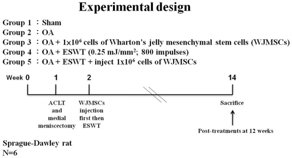
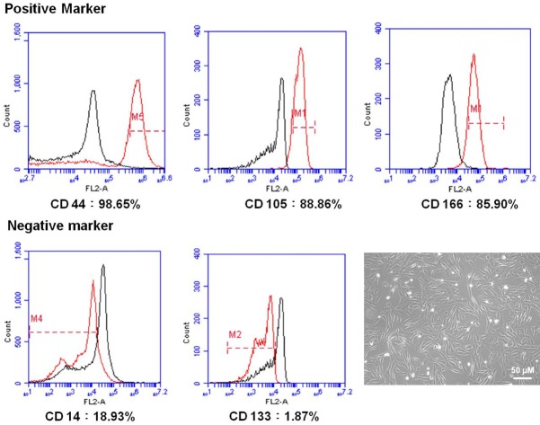
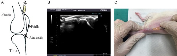
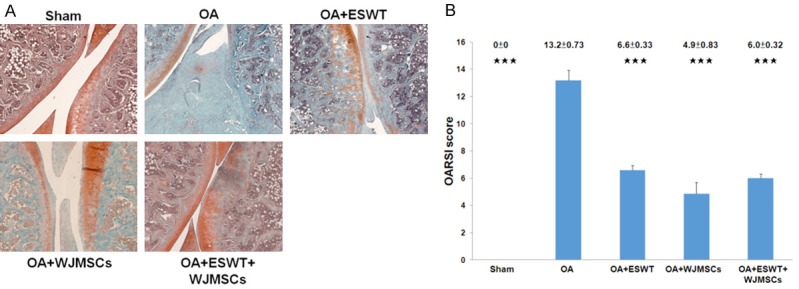
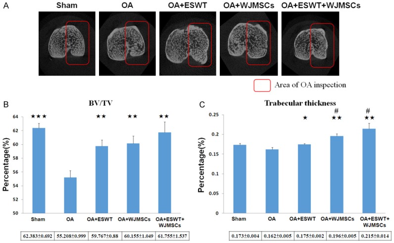
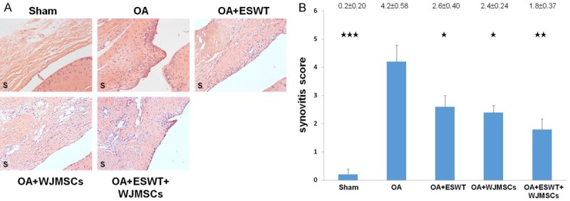
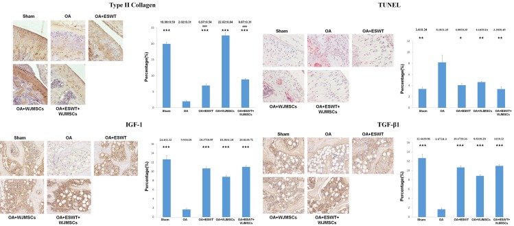
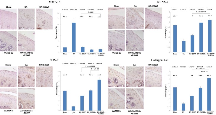
Application of extracorporeal shockwave therapy (ESWT) to the subchondral bone of medial tibia condyle has shown chondroprotective effects of the knee with decreased cartilage degradation and improved subchondral bone remodeling in the osteoarthritis (OA) of rat knee. Recently, transplantation of ex vivo preparations of mesenchymal stem cells (MSCs) to animal or human joints with OA seems to induce therapeutically effective repair because of paracrine responses from host cells including progenitor cells residing within the synovium. This study compared ESWT, Wharton's jelly mesenchymal stem cells (WJMSCs) and combination of ESWT and WJMSCs therapies for early OA of the rat knee. The results showed ESWT, WJMSCs and combination of therapies significantly improved early OA knee based on analysis of pathological findings, micro-CT and immunohistochemistry (IHC) stain. The combined therapy group increased the bone volume (61.755 ± 1.537), and trabecular thickness (0.215 ± 0.014; P < 0.01) as well as reduced synovitis (1.8 ± 0.37) more than ESWT or WJMSCs individually. However, there were no significant difference in combined ESWT and WJMSCS as shown in the expressions of IGF-1 and TGF-β1 and reduction of the TUNEL activity on OA knee. Furthermore, WJMSCs treatment significantly increased the expression of the type II collagen (22.62 ± 0.84; P < 0.001) when compared with ESWT (6.97 ± 0.54) and ESWT combined with WJMSCs (8.87 ± 0.31) in OA knee. In mechanistic factors analysis, the synergistic effect was observed by ESWT combined with WJMSCs in the expression of RUNX-2, SOX-9 and Collagen Xα1 on OA knee. Our results provided the innovative information of ESWT, and WJMSCs in the treatment of early osteoarthritis of the knee in rats.

Keywords: Shockwave therapy; Wharton’s jelly mesenchymal stem cell; articular cartilage; osteoarthritis; subchondral bone.


Conflict of interest statement

None.

Figures

Figure 1
Figure 1
The study design. Graphic scheme depicted the study design of the experiment including knee surgery, shockwave application, WJMSCs injection, and sacrifice of animals. The six rats were used in the experiments.
Figure 2
Figure 2
Characterization of WJMSCs and analysis of cell surface makers on WJMSCs. The cells were labeled with indicated markers as following: the CD44 (98.65%), CD105 (88.85%), and CD166 (85.90%) for the positive markers as well as CD14 (18.93%), and CD133 (1.87%) for the negative markers. The cultured WJMSCs showed fibroblast-like morphology.
Figure 3
Figure 3
The injection of WJMSCs. A. The knee sketch outlined the locations of WJMSCs injection in the rats knee. B. The image showed that ultrasound machine guide the localizations of WJMSCs for injection. C. The WJMSCs were injected in the knee of rats using above technique.
Figure 4
Figure 4
The pathological changes of the rat knee after treatments. A. The microphotographs of the knee showed cartilage degradation in the different groups. The microphotographs of cartilage and subchondral bone were shown the changes of cartilage in OA group, OA+ESWT, OA+WJMSCs and OA+ESWT+WJMSCs groups (n = 6). The field of the view was 50 × magnification. B. The bar chart illustrated the results of OARSI score. ***P < 0.001 as compared with OA group.
Figure 5
Figure 5
Micro-CT analysis in the proximal tibia after treatments. (A) The photomicrographs of the knee were shown in the transverse plane. The medial compartment of subchondral bone from each group was marked in red box. (B) The graphic illustrated the percentage of the trabecular bone volume fraction (BV/TV), and (C) trabecular thickness compared with OA groups. *P < 0.05, **P < 0.01, ***P < 0.001 comparing to OA group. #P < 0.05 was OA+WJMSCs and OA+ESWT+WJMSCs groups comparing to OA+ESWT groups. All rats were n = 6.
Figure 6
Figure 6
Evaluation of the histological changes in the membrane of OA knee before and after treatments. A. The sections of synovium were observed by HE stain at magnifications of 50 ×. B. The synovitis scores were calculated for each group in rat knee and all groups compared with OA group after treatment. *P < 0.05, **P < 0.01, ***P < 0.001. All rats were n = 6.
Figure 7
Figure 7
Immunohistochemical analysis of the specific molecular factors in the treatments on OA knee. The immunohistochemical stains (left) and quantification (right) showed the expressions of the type II collagen, TUNEL activity, IGF-1 and TGF-β1 after treatments on OA knee. The type II collagen and TUNEL activity were measured in the articular cartilage as well as IGF-1 and TGF-β1 were observed in the subchondral bone of the knee. The field of view was 100 × magnification. *P < 0.05, **P < 0.01, ***P < 0.001 comparing to OA group. The ###P < 0.001 comparing to OA+WJMSCs group. All rats were n = 6.
Figure 8
Figure 8
The expression of the factors in the chondrogenesis after treatments on OA knee. The immunohistochemical stains (left) and quantification (right) showed the levels of the MMP-13, RUNX-2, SOX-9 and Collagen Xα1 after treatments on OA knee. The field of view was 100 × magnification. *P < 0.05, **P < 0.01, ***P < 0.001 comparing to OA group. The #P < 0.05, ##P < 0.01 and ###P < 0.001 comparing to OA+ESWT group. All rats were n = 6.

References

    1. Wang CJ, Weng LH, Ko JY, Wang JW, Chen JM, Sun YC, Yang YJ. Extracorporeal shockwave shows regression of osteoarthritis of the knee in rats. J Surg Res. 2011;171:601–608. - PubMed
    1. Wang CJ, Sun YC, Wong T, Hsu SL, Chou WY, Chang HW. Extracorporeal shockwave therapy shows time-dependent chondroprotective effects in osteoarthritis of the knee in rats. J Surg Res. 2012;178:196–205. - PubMed
    1. Kamelger F, Oehlbauer M, Piza-Katzer H, Meirer R. Extracorporeal shock wave treatment in ischemic tissues: what is the appropriate number of shock wave impulses? J Reconstr Microsurg. 2010;26:117–121. - PubMed
    1. Zhang X, Yan X, Wang C, Tang T, Chai Y. The dose-effect relationship in extracorporeal shock wave therapy: the optimal parameter for extracorporeal shock wave therapy. J Surg Res. 2014;186:484–492. - PubMed
    1. Rompe JD, Kirkpatrick CJ, Kullmer K, Schwitalle M, Krischek O. Dose-related effects of shock waves on rabbit tendo Achillis. A sonographic and histological study. J Bone Joint Surg Br. 1998;80:546–552. - PubMed

LinkOut - more resources