. 2019 Feb 15;11(2):624-640.
eCollection 2019.

Loss of Jak2 protects cardiac allografts from chronic rejection by attenuating Th1 response along with increased regulatory T cells

Affiliations

Loss of Jak2 protects cardiac allografts from chronic rejection by attenuating Th1 response along with increased regulatory T cells

Hassan Mohammed Khair Ibrahim Higazi et al. Am J Transl Res. .

Abstract

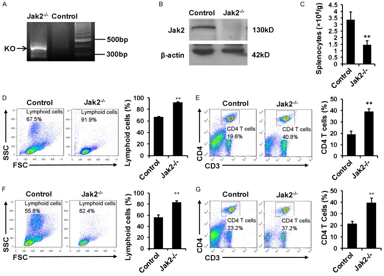
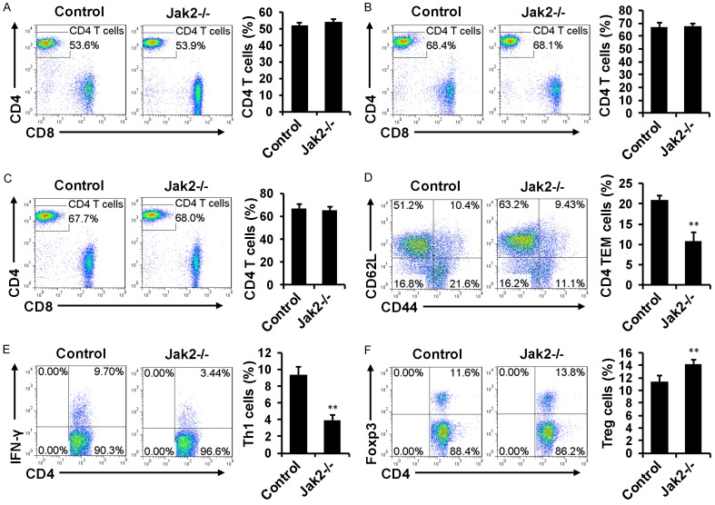
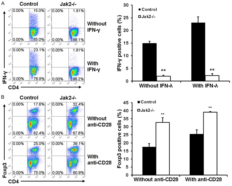
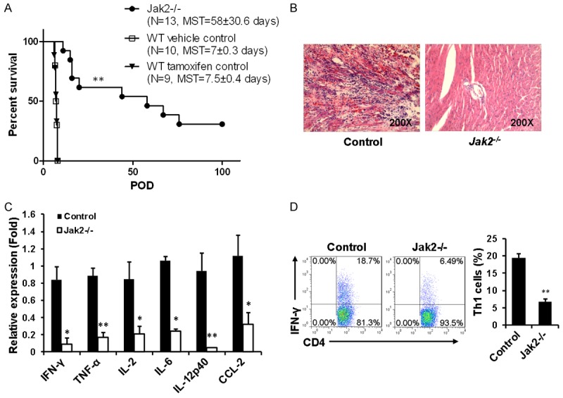
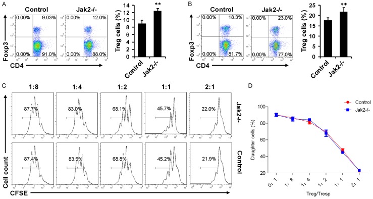
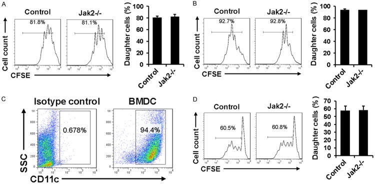
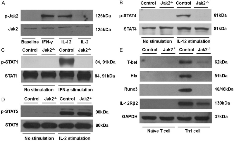
Chronic rejection acts as the most formidable obstacle for organ transplantation in clinical settings. Herein we demonstrated in a cardiac transplantation model that blockade of Janus kinase 2 (Jak2) provides protection for cardiac allografts against chronic rejection. Specifically, loss of Jak2 almost completely abolished the production of IFN-γ+ Th1 cells, while the percentage of Foxp3+ regulatory T cells (Tregs) was significantly increased. As a result, loss of Jak2 significantly prolonged allograft survival (58 ± 30.6 days vs. 7 ± 0.3 days). Particularly, 4 out of 13 Jak2 deficient recipients (30%) showed long-term acceptance of allografts as manifested by the graft survival time > 100 days. Cellular studies revealed that Jak2 deficiency did not impact the intrinsic proliferative capability for CD4+ T cells in response to nonspecific polyclonal and allogenic stimulation. Mechanistic studies documented that the impaired Th1 development was caused by the attenuated IFN-γ/STAT1 and IL-12/STAT4 signaling along with repressed expression of Th1 transcription factors T-bet, Hlx and Runx3. However, the IL-2/STAT5 signaling remained intact, which ensured normal Treg development in Jak2-/- naïve CD4 T cells. Together, our data support that blockade of Jak2 may have therapeutic potential for prevention and treatment of allograft rejection in clinical settings.

Keywords: Jak2; allograft; cardiac transplantation; chronic rejection; regulatory T cells.


Conflict of interest statement

None.

Figures

Figure 1
Figure 1
The impact of Jak2 depletion on T cell development. Jak2 deficiency was induced by tamoxifen injection. Four days after last induction, the mice were sacrificed and used for following experiments. A. PCR analysis of tail genomic DNA to check the presence of floxed null allele. B. Western blot analysis to confirm Jak2 depletion in splenic cell lysates. C. Comparison of total splenic cell numbers between Jak2-/- and control mice. Relative cell numbers were normalized by body weight. D. Flow cytometry analysis of splenic cell populations by plots on forward scatter and side scatter. Comparison was carried out for the lymphoid cluster (lower left corner) between Jak2-/- and WT controls. E. Flow cytometry analysis for the percentage of CD3+CD4+ T cells in total splenocytes. F. Flow cytometry analysis of lymphoid cells in peripheral blood. The number of lymphoid cells (lower left corner) was compared between Jak2-/- and WT controls. G. Comparison for the proportion of CD3+CD4+ T cells in peripheral blood mononuclear cells (PBMCs) between Jak2-/- and WT controls. All data were expressed as means ± SD, and four mice were included in each study group.
Figure 2
Figure 2
The impact of Jak2 deficiency on CD4+ T cell development. Loss of Jak2 did not affect the percentage of CD4+ T cells in total CD3+ splenic cells (A), total CD3+ lymph node cells (B), and total CD3+ PBMCs (C). However, the percentage for CD4+CD44highCD62Llow effector/memory T cells (TEM cells) in total CD4+ splenocytes (gated on CD4+ cells) was significantly reduced (D), and similarly, a significant reduction for the percentage of IFN-γ+ Th1 cells was noted (E). On the contrary, a significant increase for the proportion of Foxp3+ Treg cells in CD4+ splenocytes was characterized (F). All flow cytometry data were expressed as mean ± SD, and four mice were analyzed for each group.
Figure 3
Figure 3
The effect of Jak2 deficiency on Th1 and Treg development. CD4+CD62LhighCD44low naïve CD4+ T cells were purified from Jak2-/- and control mice by magnetic beads as described (cell purity > 85%). A. Loss of Jak2 impaired Th1 development. Naïve CD4+ T cells were cultured under Th1 condition in the presence (lower) or absence (upper) of IFN-γ (50 ng/ml) for five days. The production of IFN-γ secreting Th1 cells were estimated by intracellular staining followed by flow cytometry analysis. B. Loss of Jak2 enhanced Treg production. Naïve CD4+ T cells were induced under Treg condition in the presence (lower) or absence (upper) of anti-CD28 (1 ug/ml) for five days. The production of Foxp3+ Tregs was estimated by flow cytometry as above. Three mice were analyzed in each study group, and the studies were conducted with three replications.
Figure 4
Figure 4
Cardiac allografts were protected from chronic rejection in recipients deficient in Jak2. Allogenic hearts originated from BALB/c (H-2d) mice were implanted into Jak2-/- (H-2b) recipients (n=13), corn oil induced control recipients (H-2b, n=10), and tamoxifen induced Cre-ERT2 recipients (H-2b, n=9) as described, respectively. A. Allograft survival curve generated by the Kaplan and Meier method. B. Histological analysis of allograft sections six days after transplantation. C. Real-time PCR analysis of inflammatory cytokines and chemokines within the grafts six days after transplantation. D. Comparison of splenic IFN-γ+ Th1 cells between Jak2-/- and corn oil induced control recipients six days after transplantation. Four mice were included in each group for the above studies.
Figure 5
Figure 5
Comparison of Treg suppressive function between Jak2-/- and control recipients six days after transplantation. A. Comparison of splenic Treg numbers between Jak2-/- and control recipients. B. Analysis of Tregs in the draining lymph nodes (DLN) of recipients. C. A representative result for Treg suppressive assays. Proliferation of Tresps upon anti-CD3 stimulation in the presence of accessory cells along with different proportion of Tregs was estimated by flow cytometry based on the halving of CFSE fluorescence after three days of culture. D. Results for Treg suppressive kinetics of all recipients studied for each group. Four recipients were included for each group and the studies were carried out with three replications.
Figure 6
Figure 6
Analysis of the intrinsic proliferative capability of CD4+ T cells after Jak2 depletion. CD4+ T cells were prepared from Jak2-/- and control mice and labeled with CFSE to serve as responder cells, while T cell depleted control splenocytes were treated with mitomycin C to serve as accessory cells. A. Results for proliferation of responder cells stimulated with PMA (10 ng/ml) and Ionomycin (250 ng/ml). B. Proliferation results for responder cells stimulated by anti-CD3 (0.5 ug/ml) and anti-CD28 (0.5 ug/ml). C. Flow cytometry analysis of allogenic BMDCs prepared from BALB/c mice. D. Proliferation results for responder cells stimulated by BALB/c-derived allogenic BMDCs. Mitomycin C treated BMDCs were co-cultured with CFSE labeled splenic cells originated from Jak2-/- or control mice for 72 h. Cell proliferation was estimated as above based on the halving of CFSE fluorescence intensity. Similarly, four mice were included for each group and the studies were carried out with three replications.
Figure 7
Figure 7
Loss of Jak2 selectively repressed signals essential for Th1 development. A. IFN-γ and IL-12 were potent to stimulate Jak2 phosphorylation (p-Jak2) in CD4+ T cells, while p-Jak2 was undetectable in IL-2 stimulated CD4+ T cells. B. Loss of Jak2 in CD4+ T cells impaired IL-12 induced STAT4 activation (p-STAT4). C. Jak2-/- CD4+ T cells manifested impaired IFN-γ/STAT1 signaling. D. Jak2 deficiency did not impact IL-2/STAT5 signaling in CD4+ T cells. E. Western blot results for analysis of transcription factors relevant to Th1 development. Naïve CD4+ T cells originated from Jak2-/- and control mice were polarized under Th1 condition for five days, followed by analysis of T-bet, Hlx, Runx3 and IL-12Rβ2 expression levels by Western blotting. GAPDH was served as loading controls, and data shown here were a representative of three independent experiments.

Similar articles

Cited by

References

    1. Schliesser U, Streitz M, Sawitzki B. Tregs: application for solid-organ transplantation. Curr Opin Organ Transplant. 2012;17:34–41. - PubMed
    1. Newell KA, Phippard D, Turka LA. Regulatory cells and cell signatures in clinical transplantation tolerance. Curr Opin Immunol. 2011;23:655–659. - PubMed
    1. Bluestone JA. Regulatory T-cell therapy: is it ready for the clinic? Nat Rev Immunol. 2005;5:343–349. - PubMed
    1. O’Shea JJ, Plenge R. JAK and STAT signaling molecules in immunoregulation and immune-mediated disease. Immunity. 2012;36:542–550. - PMC - PubMed
    1. Kontzias A, Kotlyar A, Laurence A, Changelian P, O’Shea JJ. Jakinibs: a new class of kinase inhibitors in cancer and autoimmune disease. Curr Opin Pharmacol. 2012;12:464–470. - PMC - PubMed

LinkOut - more resources