

The value of arterial spin labelling in adults glioma grading: systematic review and meta-analysis
- PMID: 30899427
- PMCID: PMC6422184
- DOI: 10.18632/oncotarget.26674
The value of arterial spin labelling in adults glioma grading: systematic review and meta-analysis
Abstract
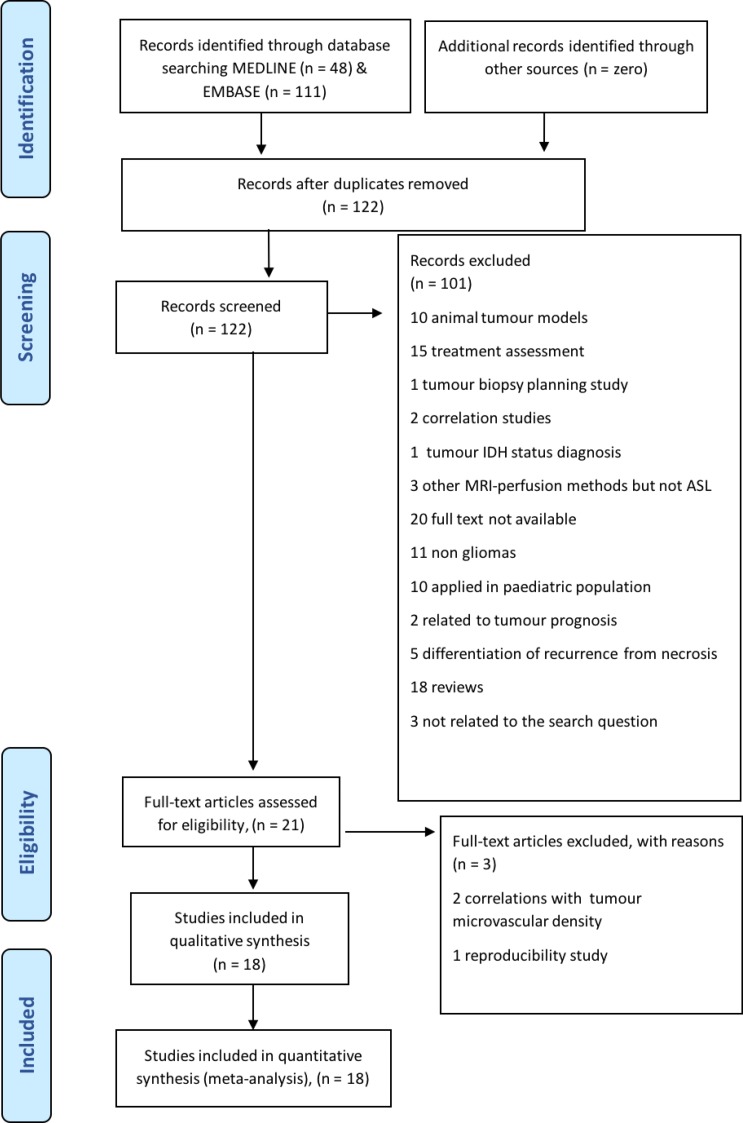
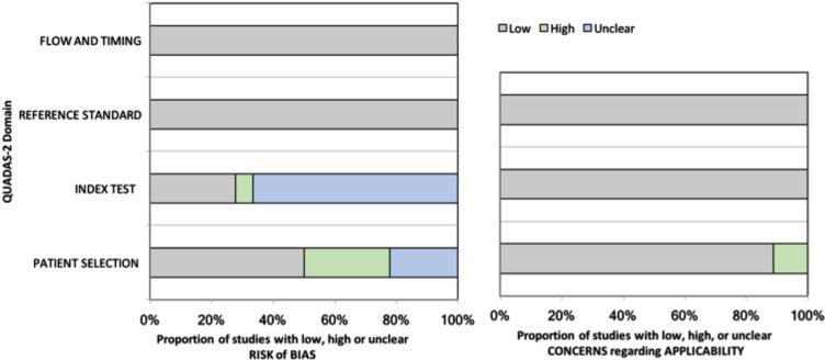
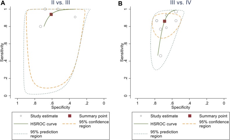
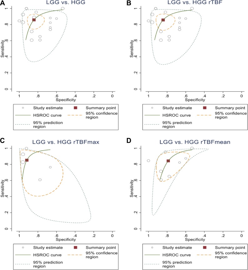
This study aimed to evaluate the diagnostic performance of arterial spin labelling (ASL) in grading of adult gliomas. Eighteen studies matched the inclusion criteria and were included after systematic searches through EMBASE and MEDLINE databases. The quality of the included studies was assessed utilizing Quality Assessment of Diagnostic Accuracy Studies-2 (QUADAS-2). The quantitative values were extracted and a meta-analysis was subsequently based on a random-effect model with forest plot and joint sensitivity and specificity modelling. Hierarchical summary receiver operating characteristic (HROC) curve analysis was also conducted. The absolute tumour blood flow (TBF) values can differentiate high-grade gliomas (HGGs) from low-grade gliomas (LGGs) and grade II from grade IV tumours. However, it lacked the capacity to differentiate grade II from grade III tumours and grade III from grade IV tumours. In contrast, the relative TBF (rTBF) is effective in differentiating HGG from LGG and in glioma grading. The maximum rTBF (rTBFmax) demonstrated the best results in glioma grading. These results were also reflected in the sensitivity/specificity analysis in which the rTBFmax showed the highest discrimination performance in glioma grading. The estimated effect size for the rTBF was approximately similar between HGGs and LGGs, and grade II and grade III tumours, (-1.46 (-2.00, -0.91), p-value < 0.001), (-1.39 (-1.89, -0.89), p-value < 0.001), respectively; while it exhibited smaller effect size between grade III and grade IV (-1.05 (-1.82, -0.27)), p < 0.05). Sensitivity and specificity analysis replicate these results as well. This meta-analysis suggests that ASL is useful for glioma grading, especially when considering the rTBFmax parameter.
Keywords: arterial spin labeling; glioma; grading.
Conflict of interest statement
CONFLICTS OF INTEREST The views expressed in this article are those of the author(s) and not necessarily those of the NHS, the NIHR, or the Department of Health and Social Care.
Figures
Similar articles
-
Efficacy of three-dimensional arterial spin labeling and how it compares against that of contrast enhanced magnetic resonance imaging in preoperative grading of brain gliomas.Environ Toxicol. 2023 Jul;38(7):1723-1731. doi: 10.1002/tox.23800. Epub 2023 Apr 11. Environ Toxicol. 2023. Retraction in: Environ Toxicol. 2025 Mar;40(3):497. doi: 10.1002/tox.24452. PMID: 37040330 Retracted.
-
A meta-analysis of arterial spin labelling perfusion values for the prediction of glioma grade.Clin Radiol. 2017 Mar;72(3):255-261. doi: 10.1016/j.crad.2016.10.016. Epub 2016 Dec 6. Clin Radiol. 2017. PMID: 27932251 Review.
-
The role of APT imaging in gliomas grading: A systematic review and meta-analysis.Eur J Radiol. 2020 Dec;133:109353. doi: 10.1016/j.ejrad.2020.109353. Epub 2020 Oct 16. Eur J Radiol. 2020. PMID: 33120241
-
Intravoxel incoherent motion diffusion-weighted imaging analysis of diffusion and microperfusion in grading gliomas and comparison with arterial spin labeling for evaluation of tumor perfusion.J Magn Reson Imaging. 2016 Sep;44(3):620-32. doi: 10.1002/jmri.25191. Epub 2016 Feb 16. J Magn Reson Imaging. 2016. PMID: 26880230
-
Glioma Grade Discrimination with MR Diffusion Kurtosis Imaging: A Meta-Analysis of Diagnostic Accuracy.Radiology. 2018 Apr;287(1):119-127. doi: 10.1148/radiol.2017171315. Epub 2017 Dec 4. Radiology. 2018. PMID: 29206593
Cited by
-
Multiparametric simultaneous hybrid 18F-fluorodeoxyglucose positron emission tomography/magnetic resonance imaging (18F-FDG PET/MRI) incorporating intratumoral and peritumoral regions for grading of glioma.Quant Imaging Med Surg. 2024 Aug 1;14(8):5665-5681. doi: 10.21037/qims-24-280. Epub 2024 Jun 11. Quant Imaging Med Surg. 2024. PMID: 39144048 Free PMC article.
-
ISMRM Open Science Initiative for Perfusion Imaging (OSIPI): ASL pipeline inventory.Magn Reson Med. 2024 May;91(5):1787-1802. doi: 10.1002/mrm.29869. Epub 2023 Oct 9. Magn Reson Med. 2024. PMID: 37811778 Free PMC article.
-
3D pCASL-perfusion in preoperative assessment of brain gliomas in large cohort of patients.Sci Rep. 2022 Feb 8;12(1):2121. doi: 10.1038/s41598-022-05992-4. Sci Rep. 2022. PMID: 35136119 Free PMC article.
-
Hemodynamic Imaging in Cerebral Diffuse Glioma-Part A: Concept, Differential Diagnosis and Tumor Grading.Cancers (Basel). 2022 Mar 10;14(6):1432. doi: 10.3390/cancers14061432. Cancers (Basel). 2022. PMID: 35326580 Free PMC article. Review.
-
Experimenting with ASL-based arterialized cerebral blood volume as a novel imaging biomarker in grading glial neoplasms.Neuroradiol J. 2023 Dec;36(6):728-735. doi: 10.1177/19714009231193163. Epub 2023 Aug 7. Neuroradiol J. 2023. PMID: 37548164 Free PMC article.
References
-
- Noguchi T, Yoshiura T, Hiwatashi A, Togao O, Yamashita K, Nagao E, Shono T, Mizoguchi M, Nagata S, Sasaki T, Suzuki SO, Iwaki T, Kobayashi K, et al. Perfusion imaging of brain tumors using arterial spin-labeling: correlation with histopathologic vascular density. AJNR Am J Neuroradiol. 2008;29:688–93. doi: 10.3174/ajnr.A0903. - DOI - PMC - PubMed
-
- Shen N, Zhao L, Jiang J, Jiang R, Su C, Zhang S, Tang X, Zhu W. Intravoxel incoherent motion diffusion-weighted imaging analysis of diffusion and microperfusion in grading gliomas and comparison with arterial spin labeling for evaluation of tumor perfusion. J Magn Reson Imaging. 2016;44:620–32. doi: 10.1002/jmri.25191. - DOI - PubMed
LinkOut - more resources
Full Text Sources
